| Jun 08, 2026 |
AI integration could help atomic force microscopy optimize scans, enhance images and interpret nanostructures without human intervention.
(Nanowerk News) A research team led by Professor Lee Jeong-hoon of the Department of Integrative Energy Engineering at Korea University, in collaboration with the teams of Professor Yoon Dae-sung of the School of Biomedical Engineering and of Professor Lee Hak-ho from Massachusetts General Hospital (MGH), a teaching hospital affiliated with Harvard Medical School, has comprehensively organized the current status and future of next-generation technology that integrates artificial intelligence (AI) with atomic force microscopy (AFM) for analyzing nanostructures autonomously.
|
|
AFM is an essential analytical tool capable of observing biological materials, such as DNA, proteins, and cells, at nanometer or sub-nanometer scale and measuring their physical properties. However, conventional AFM faces the following limitations: it is slow, requires skilled experts to interpret images manually, and can suffer from low data reproducibility. In particular, because thousands of nanostructures must be identified one by one by hand, it has been difficult to apply to large-scale biomedical research or clinical diagnostics.
|
|
To overcome these limitations, research groups around the world have been conducting AFM-AI convergence studies individually, creating a need to analyze the scattered efforts in a systematic and integrated manner to establish an organizing framework. Accordingly, at the invitation of the editors of the journal ACS Nano (“AI in Atomic Force Microscopy: Advancing Biological Nanoscale Imaging and Autonomous Discovery”), the KU-led research team published a review paper presenting an integrated framework for automating the entire AFM process with AI.
|
 |
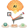
| Conceptual diagram of an autonomous bioanalysis platform using AI-augmented atomic force microscopy (AFM). AI can perform imaging-condition optimization, biomolecule prediction, modality transfer, signal interpretation, image enhancement, and structure reconstruction throughout the AFM-based measurement process. (Image: Korea University)
|
|
This study can act as a guideline that comprehensively summarizes how AI is applied across all the analytical stages of AFM, from probe optimization and scan-path planning to image denoising, super-resolution reconstruction, signal interpretation, molecular structure classification, and 3D reconstruction.
|
|
In particular, the review article analyzes how the latest deep learning techniques, such as convolutional neural networks (CNNs), which excel at image recognition and analysis; graph neural networks (GNNs), which analyze complex relational structures; generative adversarial networks (GANs), which restore image quality; and variational auto encoders (VAEs), which extract essential features from data, can address the limitations of each stage, presenting directions for future research.
|
|
The significance of this review is that it surveys how physical AI and autonomous AFM can be used in biological nanoscience. In the case of a reinforcement learning-based functional scanning system built by applying these two technologies, the scan speed, resolution, and probe force can be adjusted autonomously and in real time according to the physical properties of the sample.
|
|
This shows that AI can assist or automate the judgment processes previously performed manually by experts, demonstrating that AFM can evolve beyond a simple measurement tool into an autonomous experimental platform capable of adjusting its own measurement conditions and interpreting data.
|
|
Professor Lee Jeong-hoon said, “Physical AI-based autonomous AFM will dramatically accelerate the pace of discovery in diverse fields such as materials science, drug development, and cancer diagnosis, and we will focus on securing proprietary core technologies that lead this field.”
|