Custom small-diameter vascular grafts spun from nanofibers in minutes sustained blood flow and tissue remodeling in rats for four weeks.
(Nanowerk Spotlight) The blood vessels most often damaged in traumatic injuries are also the smallest. Arteries and veins supplying the limbs, the gut, and deep tissues measure just a few millimeters across. When one is severed, a surgeon must restore flow quickly or the downstream tissue dies. The replacement conduit must match the injured vessel’s diameter and shape closely, because even a small mismatch raises the risk of clotting or narrowing.
For larger vessels, surgeons have reliable options. Synthetic tubes made from materials like expanded polytetrafluoroethylene have served as bypasses and dialysis grafts for decades. Below about 6 mm in diameter, however, these prostheses fail at unacceptable rates. No clinically approved replacement exists for small-caliber vessels. That leaves surgeons to harvest a vein from elsewhere in the patient’s body, adding operative time and a second wound.
Filling this gap has proved difficult because each existing fabrication method solves only part of the problem. Electrospinning generates nanofiber scaffolds that resemble natural vessel walls but produces them slowly and only in simple tube shapes. 3D bioprinting recreates patient-specific geometries but cannot resolve the nanoscale structural cues that cells need to organize into functional tissue. Tissue-engineered grafts grown from living cells offer biological compatibility but require weeks to months of culture time before implantation.
A study published in Advanced Materials (“Small Diameter Vascular Grafts Made in Minutes”) reports a fabrication approach that bridges these limitations. Using Focused Rotary Jet Spinning, or FRJS, researchers produced customizable small-caliber vascular grafts with nanofiber architecture in under ten minutes, then showed they sustained blood flow and initiated tissue remodeling over four weeks in a rat model.
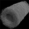
Manufacturing customizable fibrous vascular grafts. A) Fibrous vascular grafts can replace native vessels and maintain blood flow while facilitating cell infiltration and remodeling. B) (i) Excised native rat femoral artery appears as a long solid conduit, however, (ii) when decellularized, the fibrous ECM can be visualized as the base unit (scale bars: (i) 1 cm and (ii) 5 µm). C) Similarly shaped as long tubed conduits, (i) the vascular grafts appear as solid structures, (ii) also comprised of basic fibrous units on the hundreds of nanometer to single micron scale (scale bar: 5 µm). D) The vascular grafts are fabricated using FRJS, which radially extrudes a polymer jet that undergoes solvent evaporation, creating fibers which are directed toward a collector by a focused air stream. (Image: Adapted with permission from Wiley-VCH Verlag) (click on image to enlarge)
FRJS feeds a polymer solution into a reservoir spinning at up to 30 000 rotations per minute. Centrifugal force expels the solution through small orifices as thin jets. As the solvent evaporates mid-flight, polymer chains tangle into fibers that a focused air stream directs onto a rotating collection mandrel. The mandrel’s shape dictates the final graft geometry, from simple tubes to bifurcated and curved vessels, all produced as seamless continuous structures.
The polymer used was poly(L-lactide-co-ε-caprolactone), or PLCL, a biodegradable material already employed in nanofiber-based cardiovascular implants such as heart valves. At an 8% concentration in solvent, it produced fibers ranging from hundreds of nanometers to single microns. Fiber alignment varied independently of diameter and thickness, controlled by adjusting the angle between the mandrel and the air stream.
The smallest grafts, at 0.5 mm inner diameter and roughly 200 µm wall thickness, reached their final form in under 90 seconds. Larger grafts with inner diameters up to 10 mm took longer but still finished within ten minutes. These production rates exceed single-nozzle electrospinning output by orders of magnitude.
The grafts also needed to withstand physiological loads. Internal pressure testing showed they tolerated an average of 276 mmHg before fluid seeped through the porous wall, above normal blood pressure levels. No tearing or sudden rupture occurred. Suture pullout resistance measured roughly three times higher than that of native rat femoral arteries, confirming the grafts could hold surgical stitches.
Before animal studies, the team assessed whether cells could organize on and within the scaffolds. Endothelial cells seeded on the inner surface formed a continuous lining within one week, with early signs of functional cell-cell coupling at the junctions. Smooth muscle cells derived from induced pluripotent stem cells infiltrated the full wall thickness over two weeks and deposited collagen throughout.
The grafts then entered a rat femoral vasculature model as artery or vein replacements. At one week, both configurations maintained blood flow, oxygen saturation, and hemoglobin at pre-implant levels. Artery implants showed greater cell infiltration into the wall, likely driven by higher internal pressures. All animals recovered without signs of distress.
At four weeks, none of the 12 animals showed thrombosis, vessel blockage, or graft failure. Blood flow, oxygen saturation, and hemoglobin remained at or above baseline at every weekly measurement. Peripheral perfusion at the foot of the implant leg matched that of the non-implant leg, confirming adequate blood supply to downstream tissues.
Histological analysis revealed cells penetrating the full wall thickness in both artery and vein implants. Both groups showed initial endothelial layer formation along the lumen. Immune cells including macrophages and T cells had entered the graft walls, and smooth muscle organization had begun in the arterial implants. Collagen deposition appeared throughout, consistent with early-stage tissue remodeling in tubular vascular scaffolds.
Some variation appeared between samples. Certain arterial implants showed wall thinning and lumen dilation at four weeks, while others retained their original dimensions. The four-week window was too short to determine whether the immune response represents adaptive remodeling or the onset of chronic inflammation. Longer studies in larger animals with higher blood flow rates will clarify that distinction.
Several practical steps stand between these results and the operating room. Sterilization by hydrogen peroxide gas plasma takes under 30 minutes at temperatures compatible with the polymer. Residual spinning solvent can be removed by ethanol immersion in roughly ten minutes. Both timescales fit within a surgical window, but a complete intraoperative workflow has not yet been tested.
The PLCL polymer degrades over approximately one year. Matching that degradation rate to the pace of new tissue formation will be a central design challenge in future work. The FRJS platform can spin a range of natural and synthetic polymers, offering flexibility to adjust degradation profiles as the technology develops.
FRJS occupies a space between existing methods. It produces nanoscale fibers that provide cell-instructive cues, deposits them onto centimeter-scale structures of arbitrary shape, and completes the process fast enough to keep pace with surgery. The animal data confirms that these grafts can sustain vascular function and initiate biological integration over the first month after implantation. Extended studies in translational animal models will determine whether they can meet the demands of human vasculature.
For authors and communications departmentsclick to open
Lay summary
Prefilled posts
ORCID information
Kevin Kit Parker (John A. Paulson School of Engineering and Applied Sciences, Harvard University)
, 0000-0002-5968-7535 corresponding author
Nanowerk Newsletter
Get our Nanotechnology Spotlight updates to your inbox!
Thank you!
You have successfully joined our subscriber list.
Become a Spotlight guest author! Join our large and growing group of guest contributors. Have you just published a scientific paper or have other exciting developments to share with the nanotechnology community? Here is how to publish on nanowerk.com.