An origami scaffold uses electrical repair cues and light-triggered heating to promote cartilage formation while limiting vessel growth beneath the defect.
(Nanowerk Spotlight) Piezoelectric scaffolds offer a neat idea for cartilage repair: let joint movement generate electrical signals that push stem cells toward cartilage. Every step or bend could help activate the implant, turning normal mechanical loading into a repair cue. The complication is that electrical cues are broad biological signals, not instructions delivered only to cartilage-forming cells. Nearby vessel-forming cells can respond too, especially in the bone beneath the defect.
That side effect matters because the smooth cartilage lining a joint normally works without its own blood supply. If too many vessels grow beneath a repair site, they can raise oxygen levels and disturb the low-oxygen environment cartilage needs to remain stable. A scaffold that stimulates cartilage formation may therefore create a second problem unless it also limits the vascular response that stimulation can provoke.
It also contains MXene nanosheets, thin carbide flakes that heat up under near-infrared light. The folded structure strengthens the electrical response and gives cells a 3D framework to occupy, while the MXene-enabled heat restrains excessive blood vessel growth.
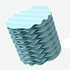
Construction of Origami-Structured PLLA/MXene Piezoelectric-Photothermal Scaffold and Its Dual Regulatory Mechanism for Cartilage Regeneration. (Image: Reproduced with permission from Wiley-VCH Verlag) (click on image to enlarge)
The design builds on wider work with biocompatible piezoelectric nanofibers, but cartilage gives the idea a sharper test. In bone repair, electrical stimulation and vessel growth can both support healing. In cartilage repair, the scaffold has to do something more selective: guide cells toward a cartilage-like matrix while preventing the repair site from becoming too vascular.
The engineering challenge was not only to make the scaffold electrically active. It also had to be porous and organized enough for cells to occupy. The researchers electrospun PLLA and MXene into fine fibrous membranes, pressed the membranes into corrugated sheets, and stacked them into a folded 3D structure. The folds concentrated stress during compression, helping the scaffold convert mechanical loading into a stronger electrical response.
With 1.5% MXene, the folded scaffold generated roughly twice the electrical output of pure PLLA under cyclic loading. That performance mattered because the scaffold’s electrical cue depends on ordinary compression rather than an implanted battery or external wiring. It also showed why the origami-like architecture was more than a shape choice. The folds helped turn mechanical deformation into a usable biological signal.
The researchers then addressed a possible false lead. MXene can conduct electricity, so a stronger signal might have reflected conductive pathways rather than true piezoelectric conversion. A control membrane with the same MXene content but randomly oriented fibers produced a far weaker response. That result pointed to aligned PLLA fibers as the main source of the motion-generated electrical cue, with MXene and folding amplifying the effect.
The scaffold also kept working under conditions closer to a joint. It maintained stronger electrical output than pure PLLA after immersion in phosphate-buffered saline and simulated synovial fluid. It degraded more slowly as well. After 12 weeks of degradation, the MXene-containing scaffold retained more mass and still produced measurable electrical output, supporting the possibility of sustained stimulation during the repair period.
MXene added the scaffold’s second control function. Under near-infrared light, the material converted optical energy into mild heat, a property also seen in near-infrared-responsive MXene materials. The 1.5% MXene scaffold reached about 41 °C in cell experiments, a temperature used to steer cell behavior rather than damage tissue. In rat joints, irradiation raised the implant-site temperature more than pure PLLA, although direct temperature measurements inside the joint remain necessary for future studies.
The effect depended on a narrow temperature range. Vessel-forming cells made more tube-like networks at about 39 °C, but those networks decreased at about 41 °C. VEGF signaling, one of the major drivers of blood vessel formation, followed the same pattern. The scaffold therefore did not use heat as a blunt tool. It used controlled mild warming to counter the vessel-promoting side of electrical stimulation.
The stem-cell experiments showed the other half of the split. Mechanical stimulation of MXene-containing scaffolds increased cartilage-related markers in bone marrow mesenchymal stem cells. Adding near-infrared stimulation strengthened that response and increased glycosaminoglycan deposition, meaning the cells produced more of the water-binding matrix molecules that help cartilage resist compression. Markers associated with fibrocartilage and unwanted mineralizing behavior did not rise in the same way.
The researchers traced the pro-cartilage effect to TRPV4, a cellular sensor that responds to both mechanical and thermal signals. When they reduced TRPV4 activity, or blocked a downstream signaling route involved in cell survival and differentiation, the cartilage-promoting response weakened. That result helped connect the material’s physical properties to a biological mechanism, rather than treating electrical and thermal stimulation as separate effects.
Blood-vessel growth followed the opposite pattern. Piezoelectric stimulation alone activated signals associated with new vessel formation. Adding mild photothermal stimulation dampened that response. The important point is the separation of outcomes. The same scaffold encouraged stem cells to make cartilage-like matrix while discouraging nearby vessel-forming cells from building vascular networks beneath the repair site.
The key test was whether that balance held up in living joints. The researchers created osteochondral defects in rat knees, meaning injuries that crossed the cartilage and reached the bone below it. They compared untreated defects, near-infrared irradiation alone, pure PLLA scaffolds, MXene scaffolds, and MXene scaffolds with irradiation. After 6 and 12 weeks, the irradiated MXene scaffold produced the most complete surface repair among the tested groups.
Tissue analysis supported the visible repair. The irradiated MXene scaffold produced stronger staining for glycosaminoglycans and type II collagen, a major collagen associated with healthy joint cartilage. The repaired region also contained more rounded cartilage-like cells rather than the elongated cells often linked to fibrocartilage. The result places the work alongside other nanomaterial strategies to guide stem cells toward bone and cartilage tissue, but with an added focus on restraining vessel growth.
The same treatment reduced vessel growth beneath the repaired cartilage. Compared with the MXene scaffold without irradiation, the irradiated scaffold showed lower vascular staining in the subchondral region and stronger markers of a low-oxygen response. Within the rat model, this did not appear to compromise repair of the underlying bone, an important point because cartilage repair still depends on stable integration with the tissue below it.
The scaffold remains far from clinical use. Rat defects do not capture the size, loading patterns, or disease complexity of human cartilage injuries. The laboratory loading protocol also simplified real joint motion, and repeated near-infrared treatment would require a practical delivery method. Future work will need to test whether the biology scales to larger joints and whether clinicians could safely control heat and electrical stimulation inside a moving joint.
The design points to a broader rule for cartilage repair: scaffolds may need to prevent the wrong kind of healing while promoting the right one. Blood vessel growth helps many injured tissues, but cartilage repair depends on keeping that response in check. By combining folded architecture, motion-generated electrical stimulation, and light-controlled heating, the PLLA MXene scaffold offers a strategy for controlling not only how much tissue forms, but what kind of tissue forms.
For authors and communications departmentsclick to open
Lay summary
Prefilled posts
Nanowerk Newsletter
Get our Nanotechnology Spotlight updates to your inbox!
Thank you!
You have successfully joined our subscriber list.
Become a Spotlight guest author! Join our large and growing group of guest contributors. Have you just published a scientific paper or have other exciting developments to share with the nanotechnology community? Here is how to publish on nanowerk.com.