| May 12, 2026 |
Nanoscale X-ray method reveals hidden orientation in materials and biological structures too small to image directly, opening new ways to study nano-order.
(Nanowerk News) Whether in tooth enamel or in nanomaterials made of silicon, the orientation of tiny internal structures often determines the properties of a material.
|
|
A new X-ray method can even make this nano-order visible when the structures are actually too small to be imaged directly. The method was developed by an international team led by the Helmholtz Centre Hereon. It opens up new possibilities to investigate materials and biological structures.
|
|
The team presents the study in the journal Light: Science & Applications (“Directional dark field for nanoscale full-field transmission X-ray microscopy”).
|
 |
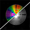
| Scattering at a test structure. The new method allows not only the location of the scattering (bottom right) to be measured, but also the orientation of the structures (top left). The orientation can be represented as color in the image. (Image: Sami Wirtensohn)
|
|
In medical X-ray imaging, the picture is created by the varying attenuation of X-rays in the body. In order to examine materials or biological tissue in detail, experts use advanced techniques that provide additional information, such as dark-field imaging. This technique exploits the fact that X-rays are scattered, i.e. deflected, at internal interfaces and irregularities.
|
|
“The scattering reveals a lot about internal structures that are not directly visible in the actual image,” explains Hereon researcher Sami Wirtensohn, first author of the study.
|
|
To make these fine structures visible, the dark field method blocks the direct X-ray beam. This allows the detector to capture only the radiation scattered inside the sample. Until now, this method has only been able to show that such structures exist, but not how they are spatially aligned. A team involving Hereon and working groups from Hamburg, Munich, Vienna, Shanghai, and Villigen (Switzerland) has now remedied this shortcoming.
|
|
“We can now also evaluate the scattering in a direction-dependent manner,” explains Wirtensohn. “This allows us to determine how the nanostructures are aligned—pixel by pixel and below the resolution limit.”
|
A simple trick with a big impact
|
|
The experimental trick is astonishingly simple and can be adapted to existing equipment with little effort: additional apertures in the beam path ensure that the sample is illuminated from different angles one after the other. By combining several images, it is possible to reconstruct how the internal structures are oriented. To test the method, the team conducted experiments at the DESY research center in Hamburg. There, the PETRA III storage ring provides intense, focused X-ray radiation. At the P05 imaging beamline operated by Hereon, the team is investigating nanoporous silicon and human tooth enamel with mineralization disorders, among other things.
|
|
This is where the strength of the method becomes apparent: the enamel nanocrystals are only a few dozen nanometers in size, yet their orientation was successfully measured.
|
 |
| Scattering on human tooth enamel with mineralization disorder (MIH). The new method enables the orientation of crystals in enamel to be determined, which is crucial for its stability and functionality. (Image: Sami Wirtensohn)
|
|
“We can see not only that something is scattering, but also how it is oriented,” explains co-author Dr Silja Flenner. “We are particularly sensitive to structures in the range of 30 to 70 nanometers – i.e., where non-destructive examination using conventional imaging techniques becomes difficult.”
|
|
The additional information about the orientation of nanostructures could be useful in several ways. In materials research, it determines how stable, conductive, or resilient a material is – for example, in porous metals, functional nanomaterials, or battery research. And in biomedicine, it provides indications as to how pathological changes manifest themselves on the nanoscale, for example in tooth enamel or bone structures.
|
|
For the Cluster of Excellence “BlueMat: Water-Driven Materials,” these findings are of central importance, as the distribution and dynamics of water in materials depend significantly on nanoscale structures. Making the finest material structures visible using the new method is crucial for the development of functional materials.
|
|
In addition, the new method provides fundamental insights for related fields of research. Only precise knowledge of structural properties down to the nanoscale makes it possible to realistically model, thoroughly understand, and specifically optimize processes in modern chemical reactors.
|