A pore smaller than one nanometer reads peptide sequences amino acid by amino acid, pinpointing single-site Alzheimer’s mutations and modifications with single-residue resolution.
(Nanowerk Spotlight) Detecting Alzheimer’s disease early means finding molecular traces of it in blood or cerebrospinal fluid, sometimes when only a few hundred copies of a telltale peptide are present among thousands of other proteins. The 42-residue fragment of amyloid-beta, known as Aβ₁₋₄₂, is one such molecule: it aggregates into the neuritic plaques that define the disease, and subtle variants of it, differing by a single amino acid substitution or a small chemical tag, may signal its onset.
But identifying those variants requires reading the peptide’s exact sequence of amino acids, building blocks packed less than half a nanometer apart, each smaller than a cubic nanometer in volume.
No existing tool does this well. Mass spectrometry, the workhorse of proteomics, fragments peptides into short pieces and reconstructs sequences indirectly through database matching. Its detection floor sits at roughly six million copies of a mid-sized protein, far too high when clinically relevant biomarkers can be present at concentrations as low as a few hundred copies per cell. Protein concentrations in human serum span 10 to 12 orders of magnitude, but mass spectrometry’s dynamic range covers only three to five, leaving the rarest molecules out of reach.
Enzyme-driven nanopore methods, which attempt to thread proteins through a biological pore one step at a time, have fared little better, reaching only 28 % to 61 % accuracy, insufficient for reliable identification, even after ten repeated passes over the same molecule.
A study published in Advanced Functional Materials (“Peptide Sequencing With Single Acid Resolution Using a Sub-Nanometer Diameter Pore”) now reports a fundamentally different strategy. Researchers at the University of Notre Dame and the University of Texas at Austin have shown that a pore less than one nanometer in diameter, drilled through an amorphous silicon membrane just a few nanometers thick, can read a peptide’s amino acid sequence one residue at a time.
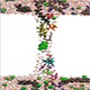
How a sub-nanometer pore reads a peptide’s amino acid sequence. Top left: an electron micrograph of the ultra-thin amorphous silicon membrane, just a few micrometers across, through which a single pore is drilled. Top center: the pore immediately after fabrication, roughly 1.5 nanometers in diameter. Top right: after exposure to air, a native oxide shrinks the pore’s narrowest point to approximately 0.4 by 0.7 nanometers, small enough to resolve individual amino acids. The line profile below the pore image shows its biconical, funnel-like shape tapering to a narrow waist. Center left: electron microscope images of single denatured Aβ₁₋₄₂ peptides coated with the detergent SDS, appearing as straight, rod-like strands less than 1.5 nanometers wide and up to 17.5 nanometers long, confirming that the treatment unfolds the peptide into a linear form suitable for threading through the pore. Center right: a computer simulation showing what a single Aβ₁₋₄₂ molecule would look like under the same imaging conditions, closely matching the experimental images. Bottom left: a molecular dynamics snapshot of an Aβ₁₋₄₂ peptide being pulled through the pore, surrounded by water molecules and sodium and chloride ions. Bottom center: a real-time recording of the ionic current as a single peptide passes through the pore. The steady baseline current drops sharply during the translocation, and fine fluctuations within that drop correspond to individual amino acids obstructing the ion flow. Bottom right: a heat map showing the distribution of hundreds of such translocation events, plotted by the depth of the current blockade versus its duration, which is used to select the most consistent events for sequence analysis. (Image: Reproduced from DOI:10.1002/adfm.202515800, CC BY)
Working with Aβ₁₋₄₂ and several variants linked to Alzheimer’s disease, the team detected a single-point mutation and a sugar-group modification at their precise positions along the chain, using only a few hundred molecules. Among the variants tested was the so-called “Arctic” mutation, a naturally occurring single amino acid substitution associated with an aggressive, early-onset form of the disease found in families in northern Sweden.
When a voltage is applied across the membrane immersed in salt solution, ions flow through the pore as a steady current. As a peptide threads through the narrowest point, each amino acid partially obstructs that flow. The size of each obstruction depends on the residue’s volume, charge, mobility, and the number of surrounding water molecules, producing a fluctuation pattern that encodes the sequence.
Fabricating a pore small enough to resolve individual amino acids, which average about 0.11 cubic nanometers in volume and sit roughly 0.38 nanometers apart along the peptide backbone, demands extraordinary control. The researchers used a focused, high-energy (300 keV) electron beam in an aberration-corrected scanning transmission electron microscope to sputter atoms from amorphous silicon membranes only 3.5 to 6.0 nanometers thick.
The resulting pore tapers to a biconical waist of approximately 0.4 by 0.7 nanometers. A native oxide that forms after air exposure stabilizes the geometry, reduces surface charge, and creates a hydrophilic lining that helps molecules pass with minimal friction.
Before entering the pore, each peptide is treated with sodium dodecyl sulfate (SDS), an anionic detergent that binds along the backbone, imparts a nearly uniform negative charge, and ensures the molecule enters the pore as a linear, string-like aggregate rather than a tangled coil. This step is essential for single-residue resolution: only when the peptide passes through in an extended configuration can each amino acid be read individually.
The resulting aggregate is less than 1.5 nanometers wide. As it enters the narrow waist, SDS molecules are stripped away, exposing individual amino acids to the ion current. The electric field there reaches about 3 million volts per centimeter, generating a force of 25 to 30 piconewtons, enough to suppress Brownian diffusion that would otherwise blur the reading. All measurements were performed at 4 to 7 °C to slow translocation further.
Early validation came from single-molecule experiments in which a biotinylated Aβ₁₋₄₂ peptide, tethered to an atomic force microscope cantilever, was threaded into a sub-nanopore and slowly extracted at 2 nanometers per second. Current fluctuations during extraction were spaced at intervals of about 0.45 nanometers, consistent with individual residues passing through. After alignment to molecular dynamics simulations using dynamic time warping, a technique that compensates for local variations in translocation speed, the correlation between measurement and simulation reached 0.98.
For practical sequencing, the team let peptides translocate freely. The ionic current was amplified over a 1.8-megahertz bandwidth and sampled at 3.6 million samples per second. To manage the resulting noise, polymer layers were laminated onto the membrane to cut parasitic capacitance, and an affinity propagation clustering algorithm grouped hundreds of translocation events by the similarity of their fluctuation patterns. Averaging the tightest cluster produced a low-noise “consensus” signal.
Decoding that consensus into a sequence required molecular dynamics simulations of each of the 20 standard amino acids passing through a model pore. Volume proved the strongest single predictor of blockade amplitude (Pearson correlation 0.77), but the radius of gyration, nearby water molecules, and residue mobility all contributed.
The researchers built a database of 1,843 human peptide sequences between 40 and 44 amino acids long from the UniProt repository, simulated each one, and compared the results to the empirical consensus. After initial ranking and alignment constrained by predicted residue mobility, the true peptide rose to rank first in every case tested.
A scrambled version of Aβ₁₋₄₂, containing the same amino acids in a different order, provided a key control. Its consensus signal was essentially uncorrelated with that of Aβ₁₋₄₂. When the aligned consensuses were subtracted and compared to the corresponding difference in simulations, the two tracked each other closely (Pearson correlation 0.93, where 1.0 would be a perfect match). The probability of such agreement arising by chance was less than one in 100,000, meaning the method can reliably tell these two peptides apart despite their identical composition. It reads sequence order, not merely which amino acids are present.
Three variants of Aβ₁₋₄₂ tested sensitivity to single-site modifications. The “Arctic mutation”, which replaces glutamic acid with the much smaller glycine at position 22 and is linked to an aggressive, early-onset form of Alzheimer’s found in Scandinavian families, produced a clear current drop at that exact site. The signal stood 2.7 standard deviations above background noise, meaning it was well outside the range of random fluctuation and easily identifiable.
A glycosylation at serine 26, attaching a bulky sugar group, generated an even more prominent peak at the correct position, 2.5 standard deviations above background. A phosphorylation at the same serine, however, was not reliably detected: the phosphate group’s volume is comparable to glycine’s, placing it near the measurement’s sensitivity floor.
Separate control experiments using block copolymers, peptides built from known repeating amino acid blocks, independently confirmed that abrupt changes in residue volume produce corresponding steps in blockade current, validating the basic measurement principle.
Systematic discrepancies between measurement and simulation highlight remaining challenges. Glycine, the smallest amino acid, was routinely overestimated relative to predictions, while larger residues such as phenylalanine and isoleucine were often underestimated. These patterns suggest that the electrical forces on ions and the behavior of water molecules inside a space this small are more complex than current computational models can fully capture.
The technique also requires denaturing each peptide into a linear form, so it cannot report on folded protein structures. Scaling to clinical use will demand reproducible fabrication of large arrays of uniform sub-nanopores, though the finding that pores with similar dimensions yielded correlated results suggests a workable manufacturing window.
By pairing extreme physical confinement with high-bandwidth electrical measurement and atomistic simulation, the work establishes that a single amino acid change or chemical modification in a peptide can be pinpointed from a sample of just a few hundred molecules, opening a path toward biomarker detection at sensitivities that current proteomic tools cannot approach.
For authors and communications departmentsclick to open
Lay summary
Prefilled posts
Nanowerk Newsletter
Get our Nanotechnology Spotlight updates to your inbox!
Thank you!
You have successfully joined our subscriber list.
Become a Spotlight guest author! Join our large and growing group of guest contributors. Have you just published a scientific paper or have other exciting developments to share with the nanotechnology community? Here is how to publish on nanowerk.com.