Nanoparticles built from red blood cell membranes and loaded with a sound-sensitive dye kill liver cancer cells when triggered by ultrasound while remaining nontoxic without activation.
(Nanowerk Spotlight) Red blood cell (RBC) membranes carry CD47, a protein that sends a molecular “don’t eat me” signal to immune cells, helping red blood cells evade immune clearance and circulate in the bloodstream for up to 120 days.
Synthetic drug carriers lack this protection. The immune system flags and destroys them within minutes. But strip a red blood cell down to its empty membrane, reshape it into a nanoscale vesicle, load it with a sound-sensitive dye, and the resulting nanoparticle inherits that biological camouflage. Trigger it with ultrasound, and it becomes a precisely targeted cancer-killing agent.
Liver cancer is where these properties matter most. Hepatocellular carcinoma, the most common form, remains difficult to treat. Surgery is invasive and depends on early detection. Chemotherapy causes off-target toxicity and, with prolonged use, drug resistance. While nanoparticle-based multimodal approaches to liver cancer have shown promise, light-based therapies that activate sensitizing agents to generate heat or toxic free radicals penetrate tissue only about 2 cm below the skin, ruling out deep organs like the liver.
Ultrasound reaches beyond 10 cm, making it a far more practical trigger. A study published in Advanced Functional Materials (“Engineering a Sonotherapeutic RBC Membrane‐Derived Nanoparticle Platform for the Treatment of Liver Cancer”) demonstrates this concept by building nanoparticles from murine red blood cell membranes, loading them with the FDA-approved dye indocyanine green (ICG), and activating them with clinical ultrasound to kill liver cancer cells.
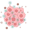
The overall concept of the developed ICG-loaded CMN for sonotherapy of liver cancer. Upon US exposure, CMN-ICG generates excessive ROS and mild temperature changes, facilitating cancer cell death and eventually restoring healthy liver function. (Image: Reproduced from DOI:10.1002/adfm.202524571, CC BY) (click on image to enlarge)
ICG is well suited as an ultrasound-activated agent. When ultrasound waves cause microbubbles in the surrounding fluid to collapse violently, the resulting flash of light, a phenomenon called sonoluminescence, excites ICG molecules into a high-energy state. As ICG returns to its resting state, it transfers energy to dissolved oxygen, spawning cytotoxic reactive oxygen species (ROS) such as singlet oxygen and hydroxyl radicals.
ICG on its own, however, performs poorly as a therapeutic. It binds rapidly to blood proteins, is cleared by the liver within minutes, and degrades quickly in aqueous solution, with a half-life of just 150 to 180 seconds.
The nanoparticle design addresses this instability by housing ICG inside vesicles made entirely from red blood cell membrane. The researchers emptied murine red blood cells through hypotonic lysis, then forced the purified membranes through successively smaller filters to produce uniform vesicles roughly 112 nm across. ICG was incubated with partially extruded membrane fragments, allowing it to associate with the phospholipid bilayer, and a final extrusion step completed the encapsulation. The resulting particles measured roughly 173 nm and achieved a loading efficiency of about 65%.
Whether the encapsulation preserved ICG’s sonodynamic function was the critical test. Spectroscopic analysis offered the first confirmation: the loaded nanoparticles retained signature absorbance peaks from both ICG and the RBC membrane proteins, indicating that the dye remained structurally intact within the carrier. In a cell-free radical-scavenging assay, the loaded nanoparticles generated ROS at levels comparable to free ICG when exposed to ultrasound at 1 W/cm², while empty membrane vesicles produced virtually none.
The nanoparticles also generated meaningful heat. Starting from physiological temperature, ultrasound exposure raised the temperature of the loaded particles to roughly 46 °C, a range associated with thermal ablation of tumor cells. This sonothermal response remained stable across three consecutive on/off ultrasound cycles, and the ICG showed no chemical degradation afterward. Together, the ROS generation and local heating represent two complementary mechanisms through which the activated nanoparticles can damage cancer cells.
Before testing anticancer effects, the researchers confirmed that the nanoparticles were safe in the absence of ultrasound. HepG2 liver cancer cells treated with the loaded nanoparticles for 24 hours showed no reduction in viability. Fluorescence imaging revealed that the particles accumulated near the nucleus after internalization, and ICG uptake was significantly higher when delivered via the membrane vesicles than as free dye, reflecting the poor aqueous stability of unencapsulated ICG.
With ultrasound, the nanoparticles shifted from inert carriers to active ones. After just 25 seconds of exposure at 1 W/cm², flow cytometry showed that roughly 42% of treated cells underwent apoptosis, compared with about 6% in cells that received the nanoparticles without ultrasound. Cell cycle analysis revealed disrupted DNA synthesis and stalled cell division.
A 30-second ultrasound treatment left only about 14% of treated cells viable, while cells exposed to ultrasound with empty membrane vesicles retained nearly 90% viability. Intracellular ROS levels rose 2.62-fold upon ultrasound activation, confirming that oxidative stress was the primary mechanism of cell death.
The researchers then moved to a three-dimensional tumor model to test whether these effects held in a more physiologically relevant setting. They embedded HepG2 spheroids in collagen gels that mimicked the stiffness of extracellular matrix surrounding actual tumors, injected the loaded nanoparticles into the spheroids, and applied ultrasound at 2 W/cm² for 30 seconds. The treated spheroids shrank by roughly 50% in mean area compared with untreated controls.
The platform requires no complex chemical synthesis or surface engineering. Membrane isolation, mechanical extrusion, and dye incubation are the only steps. Because the source material is red blood cells, the method could use a patient’s own blood to produce nanoparticles with minimal immunogenicity. In vivo studies are still needed to evaluate biodistribution, circulation lifetime, and therapeutic efficacy in tumor-bearing animal models.
RBC membrane-coated nanoparticles have, however, already been shown by other research groups to accumulate preferentially in the liver and persist in circulation far longer than conventional synthetic particles. A nanocarrier that the body recognizes as self, loaded with an FDA-approved dye, and activated by a non-invasive and widely available clinical tool offers a viable path toward treating hepatocellular carcinoma and potentially other solid tumors that light-based therapies cannot reach.
For authors and communications departmentsclick to open
Lay summary
Prefilled posts
Nanowerk Newsletter
Get our Nanotechnology Spotlight updates to your inbox!
Thank you!
You have successfully joined our subscriber list.
Become a Spotlight guest author! Join our large and growing group of guest contributors. Have you just published a scientific paper or have other exciting developments to share with the nanotechnology community? Here is how to publish on nanowerk.com.