Timed release of therapeutic molecules from a soft nanofiber-hydrogel implant supports nerve regrowth and circuit repair after spinal injury, restoring movement and bladder control in preclinical models.
(Nanowerk Spotlight) When the spinal cord is injured, the real damage often begins after the initial trauma. In the hours that follow, the body launches a series of defensive responses that create lasting barriers to repair. Immune cells flood the injury site. Inflammatory chemicals spread. Reactive oxygen compounds accumulate and begin to break down healthy tissue. Within days, scar-forming cells move in and seal off the area. What starts as a wound quickly becomes a chemically hostile, physically closed environment that blocks the regrowth of neurons and prevents reconnection across the gap.
Scientists have spent decades trying to counter these biological defenses. Some approaches try to suppress inflammation. Others attempt to stimulate axon growth or replace damaged cells. But most fall short because they focus on just one part of the injury process. The problem is not only complex, it is constantly changing. Treatments that might help during the early inflammatory phase often fail if delivered too late. And by the time structural repair becomes possible, the environment may already be too damaged to support it.
The challenge is not only about what to deliver but when and where to deliver it. That insight has begun to reshape how researchers think about neural repair. Materials that once served only as physical scaffolds are now being designed to respond to biology. With the right chemistry and structure, they can carry therapeutic signals into damaged tissue, then release them in precise patterns that align with the body’s own timeline.
That concept is central to a new study published in Advanced Materials (“Spatiotemporal Delivery of Required Facilitators for Microenvironment Remodeling Propels Neural Regeneration after Spinal Cord Injury”), where researchers created a soft, biodegradable conduit that bridges a severed spinal cord and delivers a timed sequence of molecular treatments. First it releases antioxidants to contain the early chemical damage. Then it gradually provides three growth-promoting proteins that support axon extension and circuit reconnection. The conduit itself is made from porous hydrogels and nanofibers engineered to mimic the feel of living tissue and guide neurons as they regrow.
The study brings together recent advances in material science and neurobiology into a unified system designed to intervene across multiple stages of spinal cord injury.
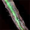
Preparation and characterization of the hybrid conduit. A) Representative digital and SEM images showing the transverse morphologies of the hybrid conduits. B) SEM images of a randomly oriented layer (left) and an uniaxially oriented layer (right) of PCL fiber mat. C) Representative digital images (GelMA (both ends),HADA/HRR (middle)) and SEM images showing the transversemorphologies of the bioactive hydrogels. D) Release profiles of CAT in the bioactive hydrogels at pH 7.4 (n = 3 per group). E) Zeta potential of peptide and growth factors. F) Release profiles of growth factor in the PCL+CAT/GelMA+EGF/NT3/GDNF/HADA/HRR hydrogel group at pH 7.4 (n = 3 per group). Data were presented as mean ± standard deviation (SD) (D–F). (Image: Reprinted with permission by Wiley-VCH Verlag) (click on image to enlarge)
In a rat model of complete spinal cord transection, this system did not simply reduce tissue damage. It shifted the biology of the wound. Neurons that would normally die survived near the lesion site. New axons extended into the damaged region. Neural circuits began to reconnect. Rats regained some ability to move, and even showed improvements in bladder function, a target rarely reached in this kind of model.
At the core of the device is a biodegradable polymer scaffold made from polycaprolactone, fabricated into a fibrous mat using electrospinning. The mat includes one layer with aligned fibers that guide regenerating axons and another with randomly oriented fibers that provide mechanical reinforcement. This scaffold wraps around a layered hydrogel structure that serves as the delivery platform for therapeutic agents.
The hydrogel is composed of two distinct sections. The outer regions contain gelatin methacryloyl, a photo-crosslinked material embedded with catalase, an antioxidant enzyme that breaks down hydrogen peroxide. This enzyme is released during the first several days after implantation, corresponding to the early inflammatory phase of the injury.
The central region of the hydrogel contains a matrix of hyaluronic acid grafted with dopamine, combined with short self-assembling peptides. This region is loaded with three key growth factors: epidermal growth factor, neurotrophin 3, and glial cell line-derived neurotrophic factor.
Each of these proteins plays a different role. Epidermal growth factor promotes the formation of extracellular matrix proteins that help support axon growth. Neurotrophin 3 regulates synaptic communication. Glial cell line-derived neurotrophic factor enhances the elongation of regenerating axons. The researchers showed that the charge interactions between these proteins and the peptide matrix influenced their release rate. Epidermal growth factor, which carries a negative charge, was released more quickly. The other two, which are positively charged, bound more strongly to the matrix and were released more gradually over two weeks.
This time-controlled release allowed the therapy to match the biological phases of injury response. Catalase was available during the acute phase to reduce oxidative damage. The growth factors followed, supporting structural and synaptic regeneration during later stages.
The device was tested in rats with a fully transected spinal cord. Compared to control animals, those treated with the full hybrid conduit showed lower levels of reactive oxygen compounds at the injury site. More neurons survived at the edges of the lesion. There was also a shift in the immune response, with a higher ratio of anti-inflammatory to pro-inflammatory markers. Markers of cell death were reduced, suggesting that the antioxidant component had a protective effect on surrounding neurons.
The researchers observed clear signs of axon regrowth. Serotonergic axons, which are critical for motor function, extended across the lesion site. These axons made contact with neurons, and the presence of synaptic proteins indicated that functional connections were being established. Electrical recordings showed that signals could once again travel across the injury site in treated animals. Behaviorally, the rats improved in joint coordination and gait. They also performed better in standardized tests of motor recovery.
Perhaps more strikingly, the treated rats showed partial restoration of bladder function. Autonomic circuits that control the bladder are particularly vulnerable to disruption after spinal cord injury and are notoriously difficult to restore. In this study, sympathetic axons marked by tyrosine hydroxylase regenerated into and beyond the lesion site. These axons formed new synaptic connections with spinal interneurons.
The researchers also observed increased expression of muscarinic acetylcholine receptors in the bladder wall, and histological analysis showed that bladder smooth muscle had recovered both in thickness and organization. Treated animals retained less urine and displayed more normal patterns of bladder function.
To trace how these regenerated axons were integrating into larger circuits, the team used a combination of viral labeling and tissue clearing techniques. They identified neurons near the lesion site that were activated and participated in signal relay across the damaged region. Some of these neurons connected with fibers originating from the brain, while others sent outputs into the lower spinal cord, forming a new bridge across the injury. These relay circuits appeared to be responsible for the restored motor and autonomic functions.
Although the treatment did not induce long-range regrowth of corticospinal axons, which are the primary pathways for voluntary movement, the formation of alternative relay circuits proved sufficient to recover some function. The study suggests that spinal repair may not require complete reconstruction of original pathways, but rather a functional reorganization that allows for signal transmission through new routes.
The work represents a shift in strategy for spinal cord repair. Rather than targeting one pathway or process, the researchers addressed multiple biological obstacles in sequence, using a material that responds to the evolving microenvironment of the injury. By aligning therapeutic delivery with the body’s own timeline, the device alters the trajectory of injury rather than attempting to reverse it outright.
While the findings are based on animal models and the system remains at a preclinical stage, the underlying design principles may apply more broadly. The combination of mechanical guidance, biochemical support, and time-controlled delivery could inform future therapies not only for spinal cord injury but also for other forms of nervous system damage where the biological response evolves over time.
For authors and communications departmentsclick to open
Lay summary
Prefilled posts
Nanowerk Newsletter
Get our Nanotechnology Spotlight updates to your inbox!
Thank you!
You have successfully joined our subscriber list.
Become a Spotlight guest author! Join our large and growing group of guest contributors. Have you just published a scientific paper or have other exciting developments to share with the nanotechnology community? Here is how to publish on nanowerk.com.