Researchers created nanoparticles that change shape inside immune cells, allowing targeted delivery of anti-inflammatory treatment and reducing joint damage in arthritis without harming healthy tissue.
(Nanowerk Spotlight) Inflamed joints, persistent pain, and progressive tissue destruction define rheumatoid arthritis, a disease where the body’s immune system turns against itself. The drivers of this destruction are well known: immune cells flood the joints and release waves of inflammatory proteins like TNF-α, setting off a cascade that damages cartilage and bone. Scientists have spent years trying to intercept this process at its source.
The logic is straightforward: stop the production of the molecules that drive inflammation, and the disease slows or even halts. One of the most direct tools for doing this is small interfering RNA (siRNA), a short stretch of synthetic genetic material designed to block specific messenger RNA molecules inside cells. But despite its precision, siRNA therapy has yet to deliver on its promise in diseases like rheumatoid arthritis. The main reason is delivery.
siRNA struggles to reach the inside of immune cells. Even when injected into the bloodstream or into tissues, it faces rapid degradation, poor cellular uptake, and most critically, entrapment in lysosomes—acidic organelles that digest foreign material. Dozens of delivery systems, from cationic lipids to viral vectors, have been developed to shuttle siRNA past these barriers. Few have succeeded in immune cells. These cells are designed to destroy what they absorb, making them among the most difficult targets for therapeutic delivery.
Strategies that work in cancer or epithelial cells often fail here. Without a way to get siRNA into the cytoplasm of immune cells safely and efficiently, its clinical utility remains limited.
A group of researchers at Zhejiang University in China now reports a delivery system that tackles this barrier with a simple but unexpected strategy: aggregation. In a study published in Advanced Functional Materials (“pH-Triggered Aggregation of Polylysine Nanoparticles Enhances TNF-𝜶 siRNA Delivery for Rheumatoid Arthritis Therapy”), they describe nanoparticles made from a modified polylysine backbone that, instead of disassembling in acidic lysosomes, do the opposite—they clump together. This acid-triggered aggregation, controlled by a chemical interaction between adamantane and cyclodextrin, gently disrupts the lysosomal membrane and allows siRNA to escape into the cytoplasm.
The result is a delivery system that enhances the potency of TNF-α–targeting siRNA in immune cells and, in a mouse model of rheumatoid arthritis, significantly reduces inflammation without detectable toxicity.
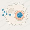
Schematic illustration of acid-triggered nanoparticle aggregation within lysosomes of immune cells, facilitating lysosomal escape, efficient TNF-𝛼 silencing and joint inflammation regulation of collagen-induced arthritis (CIA) mice. Upon lysosomal trafficking, the acidic environment (pH < 5.5) triggers the hydrolysis of the acetal groups on 𝛽-cyclodextrin (𝛽-ACD), which significantly enhances the host-guest binding affinity between 𝛽-ACD and the Ad moieties on the HBPL-Ad polymer. This aggregation causes pronounced lysosomal swelling and induces mild LMP, facilitating the escape of siRNA payloads from the lysosome into the cytosol. This hydrolysis-triggered aggregation-escape mechanism enables efficient cytosolic delivery, which is crucial for effective TNF-𝛼 gene silencing in target immune cells. This silencing, in turn, drives the strong anti-inflammatory effects in animal disease models. (Image: Reprinted with permission from Wiely-VCH Verlag) (click on image to enlarge)
The core of the delivery system is hyperbranched polylysine (HBPL), a cationic polymer with a dense network of amino groups that bind strongly to siRNA. These polymers were chemically modified with adamantane (Ad), a bulky hydrophobic molecule that can interact with cyclodextrins through host-guest chemistry—a type of selective molecular recognition. The second key component is acetalated β-cyclodextrin (β-ACD), which acts as a pH-sensitive trigger.
At neutral pH, β-ACD conceals its binding sites, preventing interaction with Ad. But when the pH drops below 5.5, as in lysosomes, the acetal groups are hydrolyzed and the exposed cyclodextrin cavities bind tightly to Ad, causing the nanoparticles to aggregate.
This design offers two advantages. First, the particles remain small and stable during circulation, which supports tissue penetration and uptake. Second, they change their structure only after reaching the lysosome. This internal aggregation is what facilitates lysosomal membrane permeabilization (LMP), allowing siRNA to escape. Unlike traditional proton sponge-based delivery strategies—which depend on creating osmotic pressure by buffering protons—this mechanism is driven by a physical restructuring of the particles in response to acid.
The researchers confirmed this mechanism with a series of chemical and physical tests. Nuclear magnetic resonance spectroscopy showed that β-ACD remains intact at pH 7.4 but undergoes rapid hydrolysis at pH 5.5, restoring the cyclodextrin’s ability to bind adamantane. Isothermal titration calorimetry demonstrated that host-guest binding between HBPL-Ad and β-ACD was negligible at neutral pH but increased sharply under acidic conditions, reaching binding constants of 10⁵ M⁻¹.
Dynamic light scattering and electron microscopy revealed that the nanoparticles grew in size and began to aggregate at lower pH, consistent with the binding data. Importantly, the siRNA remained encapsulated during this transition, and the overall particle architecture remained intact.
To test their function in cells, the researchers treated RAW264.7 macrophages, DC2.4 dendritic cells, and Jurkat T cells with fluorescently labeled siRNA packaged into these nanoparticles. The aggregated particles showed greater uptake than controls and delivered higher levels of siRNA into the cytoplasm.
In RAW264.7 cells, for example, the fluorescence intensity from internalized siRNA was more than six times higher with the pH-responsive particles compared to those delivered with Lipofectamine 2000, a widely used commercial transfection agent.
Silencing efficiency followed the same trend. After stimulation with inflammatory cytokines to induce high TNF-α expression, cells treated with the pH-responsive nanoparticles showed near-complete silencing of TNF-α mRNA. The Lipofectamine-treated cells, by comparison, achieved only partial suppression. Further imaging showed that the particles escaped the lysosome rapidly—within four hours of uptake—while Lipofectamine particles remained trapped.
Additional experiments confirmed that this escape did not rely on the proton sponge effect. Inhibition of proton pumps with bafilomycin A1 blocked the function of conventional delivery systems but had no impact on the performance of the pH-triggered aggregating particles. Acridine orange staining and time-lapse microscopy confirmed that these particles induced mild lysosomal membrane disruption, enough to release siRNA but not enough to damage the cell.
The researchers then tested the system in a collagen-induced arthritis model in mice. Mice were injected with TNF-α siRNA encapsulated in either the aggregating nanoparticles or Lipofectamine 2000. The animals receiving the pH-responsive particles showed reduced joint swelling, lower arthritis scores, and improved cartilage preservation.
TNF-α mRNA in joint tissues dropped by 93 percent, while protein levels fell by 95 percent. The corresponding reductions for Lipofectamine were 56 percent and 60 percent, respectively. Other inflammatory markers, including IL-6 and IL-1β, were also suppressed, consistent with a broader dampening of the inflammatory cascade.
Importantly, no signs of systemic toxicity were observed. Histological analysis of the liver, kidneys, spleen, and other organs showed normal structure. Blood tests indicated no disruption of liver or kidney function, suggesting that the formulation is well tolerated at therapeutic doses.
This work presents a delivery platform that is both chemically tunable and biologically precise. The approach takes advantage of a specific intracellular condition—low pH in the lysosome—to activate a structural transformation that enables endosomal escape. Unlike strategies that depend on complete disassembly or toxic triggers, this one uses aggregation as a means of escape, offering a new principle for drug delivery design.
The authors acknowledge several open questions. The precise molecular mechanism of membrane disruption by aggregation remains to be fully characterized. It is also unclear whether the same approach would be effective with other polymer scaffolds or non-cationic systems.
Additionally, while the study focused on TNF-α in rheumatoid arthritis, the platform could be adapted to deliver siRNA against other targets in other immune-driven diseases.
By solving the problem of lysosomal entrapment through an unconventional route, this study opens up a new strategy for delivering RNA therapeutics in some of the most delivery-resistant cell types. The pH-triggered aggregation of polymer-based nanoparticles offers a physically grounded, chemically specific mechanism for enhancing intracellular delivery and may inform future systems for non-viral gene therapy in inflammation and beyond, highlighting the broader potential of nanomedicine in immune-related diseases.
If this article was useful, support our independent nanotechnology reporting with any amount.
Your contribution funds the next explainer and keeps Nanowerk open for everyone.
For authors and communications departmentsclick to open
Lay summary
Prefilled posts
Nanowerk Newsletter
Get our Nanotechnology Spotlight updates to your inbox!
Thank you!
You have successfully joined our subscriber list.
Become a Spotlight guest author! Join our large and growing group of guest contributors. Have you just published a scientific paper or have other exciting developments to share with the nanotechnology community? Here is how to publish on nanowerk.com.