Researchers describe a concentric supraparticle system that protects and delivers RNA therapeutics for gene silencing in cancer models.
(Nanowerk Spotlight) In RNA-based therapeutics, scientists use RNA molecules to regulate gene expression or introduce instructions for cells to produce specific proteins. These therapies fall into two broad categories. Small interfering RNA (siRNA) is designed to silence genes by degrading messenger RNA (mRNA) molecules that carry genetic instructions from DNA. By contrast, therapeutic mRNA introduces new genetic instructions into cells, prompting them to produce beneficial proteins—such as those used in vaccines or for correcting protein deficiencies.
This approach holds promise for treating a wide range of diseases, from cancers and genetic disorders to viral infections. Unlike traditional drugs that act on proteins after they’re made, RNA therapeutics operate earlier in the biological process, potentially allowing greater control and specificity. Their modular design also allows rapid customization for different targets, which is particularly valuable in emerging infectious diseases or personalized medicine.
However, a central obstacle remains: effective delivery. Both siRNA and mRNA are fragile molecules that degrade rapidly in the bloodstream. They also struggle to reach their intended targets in the body. Once inside cells, these molecules must escape intracellular compartments and remain intact long enough to exert their effects. Many delivery platforms developed to date fall short in one or more of these areas—offering only partial protection or limited control over when and where the RNA is released.
Nanoparticle systems have offered partial solutions. Lipid nanoparticles, for instance, have been widely used for mRNA vaccines, but their structure can be unstable in the bloodstream, and repeated dosing may induce immune responses. Other carriers, such as polymeric or inorganic particles, often fail to release their payloads efficiently or degrade safely within the body.
The field has moved toward more structured and adaptive assemblies, including supraparticles—materials formed from nanoscale building blocks that organize into larger architectures with emergent properties.
Supraparticles can show distinct behaviors compared to their constituent parts. They’ve been explored in optics, catalysis, and drug delivery, particularly when arranged into regular geometries. However, their use in RNA therapeutics has remained largely undeveloped due to the challenges of constructing supraparticles that are both structurally precise and biologically functional.
A recent study published in Advanced Materials (“Self-Assembled Multilayered Concentric Supraparticle Architecture”), introduces a nanostructure that may bridge this gap. These materials, known as self-assembled multilayered supraparticles (SAMS), consist of concentric nanoscale layers formed from gold nanoparticles (AuNPs), a positively charged synthetic lipid (G0-C14), and gelatin.
The structure assembles spontaneously in water without external templating. The resulting nanospheres exhibit a layered, spherical architecture where each layer is spaced roughly 3.5 to 4.4 nanometers apart. This regularity reflects a well-defined internal organization not often achieved in nanoparticle-based carriers.
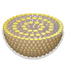
These images show how the RNA delivery particles are built and structured. Panel A illustrates the layered spherical design formed by combining gold nanoparticles, a lipid-like molecule (G0-C14), and gelatin. Panel B is a microscope image showing the layers inside the particles. Panel C confirms the regular spacing between layers using X-ray analysis, indicating a highly organized structure. (Reprinted with permission by Wiley-VCH Verlag) (click on image to enlarge)
The three components perform distinct but complementary roles. The gold nanoparticles provide a scaffold for structural integrity. The G0-C14 lipidoid, which is hydrophobic and positively charged, binds RNA molecules through electrostatic interaction. Gelatin forms a flexible outer matrix that stabilizes the system and responds to environmental cues, such as pH and enzymatic activity. The lamellar design creates confined interlayer spaces that are large enough to store small molecules like siRNA without compromising the structure.
Importantly, the self-assembly is highly sensitive to design parameters. The researchers showed that changes in nanoparticle size, lipidoid chain length, metal type, and gelatin concentration all affect the quality of the resulting SAMS. For instance, gold nanoparticles larger than 2.5 nanometers or lipidoids with excessively long or short carbon chains led to disordered assemblies.
Likewise, using proteins other than gelatin disrupted the lamellar structure. These findings highlight how the success of SAMS depends on a precise balance of chemical and physical interactions among all three components.
Molecular dynamics simulations helped clarify the mechanism behind the assembly. In particular, the presence of THPC (a stabilizing molecule bound to the gold nanoparticles) was critical. It guided the directional alignment of the G0-C14 molecules through hydrogen bonding and steric effects, creating anisotropic clustering that seeded the formation of concentric layers. Without THPC, the lipidoid chains adsorbed nonspecifically and failed to form the ordered bundles required for lamellar organization. Gelatin then anchored these assemblies, locking them into stable spheres.
This structure is not only robust during synthesis and purification—it is also responsive. Under mildly acidic conditions, which are common in tumor environments and endosomes, the particles begin to disassemble. This pH sensitivity enables the release of encapsulated RNA within target cells. Enzymes such as trypsin or MMP-2, which degrade gelatin, further trigger breakdown of the lamellae, promoting timed disassembly and intracellular release.
SAMS was tested as a delivery system for siRNA targeting the AXL gene, a known mediator of drug resistance in non-small cell lung cancer. In cultured A549 lung cancer cells, SAMS-siAXL achieved high gene knockdown efficiency, comparable to that of commercial transfection agents. Notably, this effect was achieved at lower siRNA doses, with minimal toxicity, and without the need for external transfection reagents. Imaging confirmed rapid cellular uptake, endosomal escape, and cytoplasmic release of the siRNA. The particles disassembled within 24 hours of internalization, breaking down into gold subunits small enough for renal clearance.
The platform was also tested in mouse models. Local injection of SAMS-siAXL into xenograft tumors led to reduced expression of AXL, confirming that the particles function in complex tissue environments. Systemic administration showed gene silencing effects in both conventional xenografts and patient-derived tumor models. Gene downregulation was dose-dependent and specific to the target siRNA, with no observable toxicity in major organs. Blood chemistry and tissue histology remained within normal limits, even at relatively high doses. These outcomes support the safety and reproducibility of the system in vivo.
Beyond siRNA, the authors also demonstrated that SAMS can deliver mRNA, which is larger and more structurally fragile. In a test case, the nanospheres successfully encapsulated and delivered Cre recombinase mRNA to cells, achieving gene editing comparable to standard transfection methods. This broadens the potential of SAMS to applications such as genome editing and protein replacement therapies.
The gold nanoparticles within SAMS also provide a secondary function: because gold is a strong absorber of ionizing radiation, SAMS may serve as a platform for combined gene and radiation therapies.
Compared to other delivery platforms, SAMS offers several advantages. Its architecture forms under ambient conditions without the need for organic solvents or complex fabrication steps. The concentric design provides physical protection of the payload and enables fine control over release. Because the structure is self-limiting, its size distribution is narrow, which aids consistency in dosing and biodistribution. Its disassembly is triggered by endogenous conditions, avoiding the need for external stimuli. Importantly, the components used—gold, gelatin, and lipidoids—are all familiar in biomedical research and can be scaled for clinical production.
Despite these strengths, open questions remain. The system’s performance in more complex or immunologically active environments has yet to be tested. Long-term pharmacokinetics and potential immunogenicity of the particles after repeated dosing need further study. Industrial manufacturing and quality control processes will also require refinement to maintain consistency at scale.
The study’s current scope, while thorough in design and execution, does not yet establish therapeutic efficacy beyond gene knockdown.
Nonetheless, this work presents a structured and experimentally validated system for RNA delivery that overcomes several of the key barriers faced by existing nanocarriers. The modular design allows SAMS to be adapted for different therapeutic payloads or diseases. By focusing on fundamental molecular interactions and self-organization principles, the study opens the door to a class of RNA delivery systems that are more stable, predictable, and responsive than many of their predecessors.
Get our Nanotechnology Spotlight updates to your inbox!
Thank you!
You have successfully joined our subscriber list.
Become a Spotlight guest author! Join our large and growing group of guest contributors. Have you just published a scientific paper or have other exciting developments to share with the nanotechnology community? Here is how to publish on nanowerk.com.