A physics-based model that accounts for membrane surface folds predicts cell adhesion on nanopillar arrays with 97% accuracy, enabling optimized biosensor design.
(Nanowerk Spotlight) Under a microscope, a cell’s outer surface looks nothing like the smooth bubble most people imagine. It is crumpled, ridged, and ruffled, covered in tiny folds and finger-like projections called microvilli. This extra material is not decorative. It is functional slack. When a cell needs to stretch, spread, or squeeze through a tight space, it unfurls these folds to gain additional surface area without having to manufacture new membrane or risk rupturing. Cell biologists call this stored surplus the membrane reservoir, and it plays a central role in migration, division, and wound healing.
Nanopillar arrays, surfaces covered with pillars just a few hundred nanometers tall, have become a workhorse technology in biomedical research. Depending on how deeply cells wrap around the pillars, these arrays can enhance biosensing, improve electrophysiological recording, deliver genes into cells, or extract biomolecules from them. Whether a cell perches on the pillar tips, sinks partway down, or fully encapsulates the structures dictates which of these functions the array can perform.
Experimental studies have mapped how pillar diameter, spacing, and height influence this outcome, but the results have remained qualitative. Theoretical models have failed to produce reliable predictions, largely because they treat the membrane as a simple elastic sheet and ignore the very reservoir of folded area that cells routinely deploy when adapting to new geometries.
A study published in Advanced Functional Materials (“Predicting Cell Adhesion States on Nanopillar Arrays with a Nano‐Bio Interface Model: From Modeling to Functional Device Design”) puts this reservoir at the center of a new predictive framework. The researchers developed a reservoir-guided nano-bio interface (RG-NBI) model that tracks the full adhesion process in two sequential stages. In the first, the connection between the membrane and the underlying cytoskeleton, the structural scaffolding inside the cell, stretches linearly. Tension rises in proportion to the cell’s descent into the gaps between pillars.
In the second stage, the membrane reservoir activates: folds unfurl, tension plateaus, and the cell wraps much deeper around the pillars. Whether the reservoir activates at all depends on a critical spacing threshold. Below that threshold, the geometry is too tight for enough tension to build, and the cell simply perches on the pillar tips. Above it, the reservoir engages and the cell sinks substantially further.
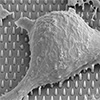
Mechanistic illustration, modeling, and functional validation of nano-bio interfaces based on NAs. (A) Schematic illustration of cell adhesion states on NAs. (B) Construction of the RG-NBI model and development of an automated analysis platform, NAs Designer, for predicting cell adhesion states. (C) Optimized NMEAs enable accurate recording of neuronal electrical activities, providing a versatile platform for mechanistic studies and therapeutic interventions in neurological disorders. (Image: Reproduced with permission from Wiley-VCH Verlag) (click on image to enlarge)
The model defines three adhesion states using a metric called the engulfment percentage, the ratio of the cell’s adhesion depth to the pillar height. A cell resting on the tips with minimal wrapping occupies the “top” state. Partial wrapping corresponds to a “middle” state, and full encapsulation of the pillars constitutes the “bottom” state. The transition from top to middle is not gradual. It is abrupt, driven by the sudden release of stored membrane area once the reservoir activates. This produces a discrete jump in adhesion depth rather than a smooth continuum.
To make the model practical, the team built a software tool called NAs Designer. Users enter three geometric parameters of a nanopillar array, namely diameter, spacing, and height, and the program outputs the predicted adhesion state within seconds. Validation against 36 experimental cases, drawn from both the team’s own work and independent published studies covering multiple cell types, yielded a prediction accuracy of 97.22%.
Separate experiments confirmed that nanopillar substrates support healthy cell growth without protein coatings. Cells cultured on nanopillar arrays achieved survival rates above 96%, comparable to collagen-coated flat surfaces and far superior to bare flat electrodes, where viability dropped to roughly 71%. This finding carries practical weight for electrode applications, because the protein coatings typically used to promote cell adhesion also shield active electrode sites and impair detection performance.
The researchers then used NAs Designer to guide the fabrication of nanopillar microelectrode arrays, electrode chips with nanopillars built directly onto the sensing sites. The design rationale followed directly from the model: electrochemical sensing benefits from maximum exposed electrode area beneath the cell, which requires a top-state configuration, while electrophysiological recording benefits from an intimate cell-electrode seal, which requires a bottom-state configuration.
For electrochemical detection of dopamine, a neurotransmitter linked to neurological disorders including Parkinson’s disease, the top-state arrays achieved a sensitivity of 353.86 nA/µM/mm² and a detection limit of 6 nM. Because cells sat on the pillar tips, most of the gold electrode surface remained exposed and available for charge-transfer reactions. On the widest-spacing arrays, by contrast, cells fully covered the electrode and eliminated virtually all active surface area.
Electrophysiological recording told the opposite story. The bottom-state arrays, where cells fully encapsulated the pillars, produced the highest signal-to-noise ratio of 7.97. The tight seal between cell and electrode reduced voltage leakage and amplified the detected signal. Without geometry-guided optimization, neither the electrochemical nor the electrophysiological configuration would have outperformed conventional planar electrodes.
The team put these optimized arrays to work by monitoring dopamine release from neuroblastoma cells stimulated with potassium chloride. The arrays captured the expected dose-dependent increase in secretion, the plateau at ion-channel saturation, and the complete suppression of release in calcium-free medium, confirming that the exocytosis pathway remained intact.
A separate experiment extended the platform to cancer metabolism by tracking choline consumption in glioblastoma cells over several days. Choline is an essential nutrient involved in membrane synthesis, and glioblastoma cells consume it at elevated rates to fuel their rapid proliferation. The arrays revealed an accelerating consumption rate that mirrored cell growth, demonstrating the platform’s utility beyond neurotransmitter detection.
By linking pillar geometry to cell adhesion through a physics-based model and packaging that model into accessible software, the study establishes a design loop in which researchers can specify a desired cell-electrode interaction and work backward to the required pillar dimensions. This provides a systematic alternative to the trial-and-error approach that has dominated nanostructured biointerface design, and it demonstrates that the membrane reservoir, long studied in the context of cell motility and shape change, is equally consequential when cells meet engineered surfaces.
For authors and communications departmentsclick to open
Lay summary
Prefilled posts
Nanowerk Newsletter
Get our Nanotechnology Spotlight updates to your inbox!
Thank you!
You have successfully joined our subscriber list.
Become a Spotlight guest author! Join our large and growing group of guest contributors. Have you just published a scientific paper or have other exciting developments to share with the nanotechnology community? Here is how to publish on nanowerk.com.