| Jun 05, 2025 |
Researchers miniaturize tumor-like environments using innovative hydrogel microfiber architectures.
|
|
(Nanowerk Spotlight) Scientists from the 3B’s Research Group at the University of Minho, in collaboration with the University of Santiago de Compostela (Spain), have developed a groundbreaking new way to mimic tumors in the lab—paving the way for faster, more accurate testing of cancer treatments. The work was led under the supervision of renowned biomaterials expert Professor Rui L. Reis.
|
|
Using a cutting-edge technique called multiphase microfluidics, the team has created hydrogel fibers that can simulate the complex conditions found inside real tumors. These 3D cancer models provide a more lifelike environment than traditional methods, making them a powerful tool for studying how tumors grow and respond to chemotherapy in a miniaturized way.
|
|
The team’s findings have been published in Matter (“Precise multiphase hydrogel engineering of miniaturized 3D cancer architectures via computationally informed microfluidics”).
|
|
“Our approach allows us to recreate the mechanical stress and nutrient limitations that cancer cells experience in the human body,” explains Dr. Ramón Rial Silva, lead researcher from the University of Santiago de Compostela. “This gives us a much clearer picture of how drugs like doxorubicin work—or fail—in real tumor conditions.”
|
|
What sets this approach apart is its combination of experimental innovation with computational modeling. Before producing the fibers in the lab, the team uses computational fluid dynamics (CFD) simulations to predict how different materials and flow conditions will affect the final structure. This allows them to fine-tune the fiber design—controlling bubble size, core thickness, and overall shape—without wasting time or lab materials. It’s a smarter, faster way to design complex biological models.
|
 |
| Overview of the multiphase microfluidic biofabrication platform developed to create miniaturized 3D cancer models. (1) Computational fluid dynamics (CFD) and solid mechanics simulations are used to predict and optimize hydrogel fiber architectures and mechanical stress conditions. (2) A custom 3D flow-focusing microfluidic chip enables continuous wet-spinning of hydrogel microfibers with embedded cancer cells. Depending on the phase composition, the system generates either air-pocket-based spheroids or continuous fiberoids, allowing the formation of structurally distinct, physiologically relevant tumor models. (Image courtey of the researchers) (click on image to enlarge)
|
|
The technology works by flowing different materials and phases—liquid, gel, and gas—through a micro-scale device to form tiny, customizable hydrogel fibers. These fibers contain either air pockets or a continuous liquid core, depending on the type of model needed. Cancer cells grow within these fibers in ways that closely mimic how they behave inside the body.
|
Two main models were studied:
• Spheroid-pocket fibers, which trap air bubbles to form small liquid chambers where cancer cells naturally form spherical clusters—called spheroids—under realistic solid stress conditions.
• Fiberoid fibers, which have a central channel filled with glioblastoma (brain cancer) cells and an outer shell containing healthy brain cells (astrocytes), allowing researchers to observe direct tumor-stroma interactions in real time.
|
|
Unlike conventional 3D cultures, this system is highly efficient and scalable. Researchers can grow hundreds of identical fiber-based tumor models with minimal handling, cutting down on time, cost, and experimental variability.
|
|
“Being able to control the size, shape, and internal environment of each fiber gives us unprecedented precision” adds Dr. Carlos F. Guimarães, second author and researcher from 3B’s Research Group, University of Minho. “It also makes it easier to test how tumors respond to different drugs, accelerating the development of more effective, personalized therapies.”
|
 |
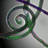
| Fluorescence microscopy image showing spiral-shaped hydrogel microfibers fabricated using the multiphase microfluidic system. The fibers contain distinct compartments marked by green, red, and blue fluorescence, representing spatially organized components such as cells and materials. This structural complexity demonstrates the platform’s ability to generate architecturally intricate and biologically functional 3D models with controlled composition and compartmentalization. Scale bar: 200 µm. (Image courtey of the researchers)
|
|
Reflecting on the broader significance of this development, Professor Rui L. Reis adds: “It’s the logical evolution to this unique way to deploy microfluidics as a biofabrication technology that we pioneered and have been developing for several years now.”
|
|
This innovation opens new possibilities not only for drug screening and mechanobiology research, but also for precision medicine, as the platform can be adapted to work with patient-derived cancer cells.
|
|
Source: Provided by the University of Minho
|
|
|
|
|