Neurons placed inside engineered living bodies built from frog cells self-organize, become active, and reshape movement without evolutionary guidance.
(Nanowerk Spotlight) Understanding how neurons organize and function inside bodies that evolution never designed matters for building functional living machines, engineering replacement tissues, and grasping how biological complexity emerges. But every neural circuit studied in nature reflects a deep co-evolutionary history in which neurons, sensory organs, and motor systems were refined together over hundreds of millions of years. Separating what neurons can do on their own from what that evolutionary context provides has remained an open experimental challenge.
Existing systems have approached pieces of the problem but none has confronted it directly. Two-dimensional neuron cultures show that neurons can develop outside an organism, but they lack three-dimensional structure and cannot move. Brain organoids develop layered circuits and spontaneous electrical activity, yet they sit inert in dishes, requiring external electronics to interact with anything. Biohybrid robots that combine living muscle with synthetic scaffolds can generate motion, but they are not fully biological and depend on external stimulation. No existing system has combined all four properties: entirely alive, self-assembling, mobile, and equipped with functional neurons.
A study published in Advanced Science (“Engineered Living Systems With Self‐Organizing Neural Networks: From Anatomy to Behavior and Gene Expression”) now fills that gap. Researchers at the Allen Discovery Center at Tufts University and the Wyss Institute for Biologically Inspired Engineering at Harvard University describe “neurobots,” autonomous living constructs assembled entirely from embryonic cells of the African clawed frog, Xenopus laevis, that contain neurons capable of self-organizing into networks within a novel, motile body.
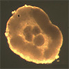
Construction and development of a neurobot. (a) Neural precursor clumps were placed in the center of an animal cap “bowl”, excised from the animal pole of a Xenopus laevis embryo before it fully closed during healing. The composite forms gradually into first a sphere and then a more elongated shape, which is mobile by Day 3. (b) Top panels: examples of two neurobots, one more rounded than the other. Bottom panel: Roundness Index (RI) was calculated by fitting an ellipse on the image of the bot and calculating the ratio between the minor and major axes. Neurobots tended to be less rounded than biobots (Kruskal–Wallis test, p = 0.047). (c) Neurobots were significantly larger than biobots (Kruskal–Wallis test, p = 0.0007). The central line on the box plot shows the median, and the bottom and top edges of the box indicate the 25th and 75th percentiles, respectively. The whiskers show the extent of the extreme data points not considered outliers, and the outliers are shown using the ‘+’ symbol. (Image: Reproduced from DOI:10.1002/advs.202508967, CC BY) (click on image to enlarge)
The neurobots build on a simpler precursor called a “biobot.” When a small patch of ectodermal tissue is cut from the outer surface of an early-stage frog embryo and left to develop on its own, it heals into a tiny sphere whose surface differentiates into four cell types normally found on tadpole skin. Among these are multiciliated cells whose rhythmic ciliary beating propels the construct through water and small secretory cells that release serotonin. These biobots are autonomous, self-powered, and survive roughly 9 to 10 days on inherited yolk reserves.
To create neurobots, the team exploited a well-established property of frog embryonic cells: when cells from the animal cap of a Xenopus embryo are dissociated and kept separated for 3 or more hours, they spontaneously adopt a neural fate. The researchers dissociated caps from approximately 50 embryos, allowed the cells to become neural precursors, then reaggregated them into small clumps. During the brief window before a freshly excised animal cap healed shut, they placed these clumps inside the closing tissue. Within 30 minutes the composite sealed itself into a sphere. By the third day, cilia appeared on the surface and the neurobot began to move.
Antibody staining confirmed that the implanted precursors had matured into neurons. These neurons extended projections both internally, among one another, and outward toward the surface, reaching the cilia-bearing cells responsible for locomotion. Additional markers revealed distinct axonal and dendritic compartments, the two fundamental types of neuronal extensions, as well as clusters of synaptic vesicles, the tiny packages neurons use to communicate at connection points.
Every neurobot developed a unique neural architecture, a consequence of the variability inherent in manual assembly. Yet a common structural feature emerged: a largely cell-free central cavity through which neurites extended, sometimes in notably straight lines, suggesting the presence of an internal support scaffold possibly composed of extracellular matrix proteins.
Calcium imaging, a technique that makes active neurons glow, confirmed that these self-organized networks were functional. In freely moving neurobots, the implanted cells produced spontaneous calcium signals. Occasionally, regions separated by considerable distance fired together, a possible sign of connectivity, though the researchers caution this could also reflect coincidence.
The behavioral consequences proved measurable. Over 30-minute observation sessions involving 47 neurobots and 48 biobots, neurobots were significantly less likely to sit idle: their minimum movement speed was notably higher. More importantly, their trajectories were more complex. While some biobots traced simple circular paths with constant radii, neurobots frequently generated elaborate patterns that varied over time.
The researchers quantified this using spectral analysis of the position data, counting unique frequency peaks across both spatial dimensions to produce a Complexity Index. A value of one indicates a simple circle; higher values reflect more intricate movement. Neurobots scored significantly higher (p = 0.039), and this difference could not be attributed to their larger size or more elongated shape. Sham neurobots, which received implanted cells reaggregated too quickly to adopt a neural fate, did not show the same gains.
Drug experiments provided additional evidence for a neural contribution to behavior. When both construct types were exposed to 15 mM pentylenetetrazole, or PTZ, a compound that blocks inhibitory GABA_A receptors and is widely used to induce seizures in animal models, biobots and neurobots responded in opposite directions. Nearly all biobots decreased their movement complexity, likely because PTZ acted on GABA receptors found on non-neuronal goblet cells, potentially altering mucus secretion and indirectly affecting ciliary motion.
Most neurobots, by contrast, increased their complexity, suggesting that neural activity actively counteracted the drug’s default suppressive effect. The difference between the two groups was statistically significant (p = 0.01) and vanished in control experiments where the drug was replaced with plain media. Neurobots raised in zolmitriptan, a serotonin receptor agonist known to promote ectopic neural growth, showed increased innervation and a tendency toward greater complexity under PTZ, hinting that serotonergic signaling during development may bias neural expression toward GABAergic subtypes.
Bulk RNA sequencing revealed a gene-expression landscape substantially different from that of plain biobots. Neurobots showed significant upregulation of 6,774 genes. The most prominent categories involved nervous system development, synapse formation, and receptors for all major neurotransmitter systems. Genes encoding voltage-gated ion channels, growth factors, and proteins important for synaptic plasticity were also highly expressed.
Among the transcriptomic results, a large cluster of upregulated genes encoded proteins involved in visual perception, phototransduction, and photoreceptor development. These included red and violet cone opsins, rhodopsin, melanopsin, and retinal G-protein coupled receptors, genes normally active exclusively in frog eyes. Genes characteristic of major retinal cell types, including retinal ganglion cells and horizontal cells, were also present.
Transcript upregulation does not guarantee protein production, and the researchers emphasize that follow-up proteomic studies will be needed to determine whether these genes translate into functional light-sensing machinery. If they do, light-responsive behavior in neurobots would represent an entirely novel emergent property with no evolutionary precedent.
Gene-expression variability across neurobot samples was significantly greater than in either biobots or shams, a pattern the authors attribute partly to the influence of neural tissue on how cells explore their genetic landscape. A phylostratigraphic analysis, which assigns genes to evolutionary age categories, revealed that over 54 percent of upregulated genes in neurobots belonged to the two most ancient categories: genes shared across all cellular organisms including bacteria, and genes common to all eukaryotes. Very few ancient genes were downregulated. Assembling neurons into a body that evolution never designed appears to push the construct’s gene-expression profile toward the distant evolutionary past.
The neurobot platform remains in its early stages. Manual construction limits reproducibility, and the constructs’ rotational movements have so far prevented researchers from directly correlating specific neural firing patterns with specific behaviors. Automation of assembly, optogenetic tools for selectively activating neuron populations, and direct tests of light sensitivity are among the planned next steps.
What the work establishes is that wild-type neurons placed in an entirely unfamiliar body can mature, wire themselves together, form synaptic structures, become spontaneously active, and measurably alter the behavior of the construct they inhabit. These neurons also activate an ancient gene-expression program, including one for sensing light. The findings suggest that the biological hardware for building a nervous system carries far more intrinsic organizational capability than any single evolutionary lineage has yet put to use.
For authors and communications departmentsclick to open
Lay summary
Prefilled posts
Nanowerk Newsletter
Get our Nanotechnology Spotlight updates to your inbox!
Thank you!
You have successfully joined our subscriber list.
Become a Spotlight guest author! Join our large and growing group of guest contributors. Have you just published a scientific paper or have other exciting developments to share with the nanotechnology community? Here is how to publish on nanowerk.com.