Therapeutic mRNA delivered via lipid nanoparticles restored sperm production in genetically infertile mice and produced live offspring without genomic integration.
(Nanowerk Spotlight) Male infertility accounts for roughly half of all infertility cases worldwide, yet treatment options for its most severe forms remain limited. Among the hardest cases to address are those caused by genetic mutations that halt sperm development partway through, a condition known as maturation arrest. In these men, germ cells begin their journey toward becoming sperm but stall during meiosis, the specialized cell division that produces cells with half the normal chromosome count. The result is non-obstructive azoospermia: a complete absence of sperm in the ejaculate, with no physical blockage to blame.
Hormone therapies rarely help these patients. Surgical sperm retrieval succeeds only in a small fraction of cases. And while gene-editing tools like CRISPR/Cas9 have corrected such mutations in animal models, they carry serious concerns. Off-target edits could introduce new mutations. More fundamentally, editing the human germline (eggs, sperm, and embryos) is ethically prohibited in most jurisdictions. Viral delivery vehicles such as adeno-associated viruses bring their own problems: they can integrate into the genome at low frequencies, provoke immune responses, and carry only limited genetic cargo.
Against this backdrop, mRNA-based therapies offer a different logic. Rather than permanently rewriting DNA, synthetic mRNA enters a cell, gets translated into the needed protein, then degrades. The protein does its job; the genome stays untouched. This principle powered the COVID-19 vaccines that reached billions of people, validating lipid nanoparticles (LNPs), fat-based nanocarriers, as safe and effective delivery vehicles for mRNA.
Researchers have since steered LNPs toward the lungs, brain, and placenta. But directing them into the testes, and specifically into the spermatocytes where meiosis occurs, had remained largely unexplored.
A study published in Advanced Science (“Testicular mRNA‐LNP Delivery: A Novel Therapy for Genetic Spermatogenic Disorders”) addresses that gap. A team based primarily at Shanghai Jiao Tong University School of Medicine, with collaborators at the Southern University of Science and Technology in Shenzhen and Nanjing Medical University, reports an LNP formulation that preferentially targets spermatocytes in living mice. The researchers used this vehicle to deliver therapeutic mRNA directly into the seminiferous tubules, the coiled tubes inside the testis where sperm develop. The treatment restored sperm production in two different mouse models of genetic meiotic arrest and produced a live offspring from the rescued sperm.
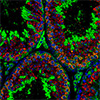
Schematic diagram of novel therapy for genetic spermatogenic disorder via testicular mRNA-LNP delivery. (Image: Reproduced from DOI:10.1002/advs.202509855, CC BY) (click on image to enlarge)
The work began with a systematic screen. The team synthesized 30 LNP formulations, each built around a different ionizable lipid, a fat molecule that changes its electrical charge depending on acidity, allowing it to escape cellular compartments after uptake. All 30 carried mRNA encoding a fluorescent reporter protein and were grouped into three pools of ten.
Each pool was injected into the seminiferous tubules of adult mice through the rete testis, a network of channels at the back of the organ. After 24 hours, the researchers checked which cell types glowed green. Only one pool delivered its cargo preferentially to germ cells rather than to surrounding support cells. Further testing identified a single formulation, designated Pool1-LNP3, that targeted spermatocytes specifically. Imaging confirmed that protein expression persisted for about seven days.
The team then turned to a disease model. Mice homozygous for a point mutation in the Msh5 gene (Msh5-D486Y/D486Y), originally identified in men with non-obstructive azoospermia, exhibit complete meiotic arrest. Their spermatocytes cannot properly repair DNA double-strand breaks or form the crossover structures essential for chromosome sorting during meiosis. No sperm are produced.
The researchers loaded wild-type Msh5 mRNA into LNP3 and injected it into the rete testis of mutant males. By day 14, round spermatids, cells that have completed meiosis, appeared in the seminiferous tubules. By day 21, elongated spermatids with normal shape were present.
Flow cytometry showed that 55.9 % of testicular cells in treated mice were haploid, compared with essentially none in untreated mutants. About 32 % of seminiferous tubule cross-sections contained spermatids with intact acrosomes, the cap-like structures sperm need for fertilization.
The effect was transient, consistent with the temporary nature of mRNA expression. Elongated spermatids appeared around day 18 and disappeared by day 22. This narrow but sufficient window fits the underlying biology: once mRNA-derived MSH5 protein enables a spermatocyte to complete meiosis, the cell proceeds through later development stages without needing continued protein supply.
A central question was whether the delivered mRNA had integrated into the genome of the rescued sperm. Sanger sequencing of ten individually isolated sperm confirmed that every one still carried the homozygous Msh5 mutation. No genomic integration had occurred.
To test fertility, the team used intracytoplasmic sperm injection (ICSI), a technique in which a single sperm head is injected directly into an egg. Of 53 injected wild-type oocytes, 30 developed to the blastocyst stage. All ten blastocysts selected for genotyping were heterozygous for the mutation, exactly as expected when a mutant sperm fertilizes a wild-type egg. After embryo transfer into surrogate mothers, a live pup was born.
The team tested the platform’s versatility in a second model: mice lacking the Maps gene, which also show spermatogenic arrest at the spermatocyte stage. Delivery of Maps mRNA via LNP3 similarly rescued sperm production, with about 29.5 % of tubule cross-sections containing spermatids by day 21.
Safety assessments showed no adverse signals. Western blots detected the reporter protein only in the testis, not in the heart, liver, spleen, lungs, kidneys, or brain. Histological examination of those organs revealed no tissue damage, and blood levels of the inflammatory marker IL-1β did not rise after injection. The blood-testis barrier, a tight-junction network that seals the seminiferous tubule interior from the bloodstream, likely helped confine the nanoparticles to their target.
The study carries limitations the authors openly acknowledge. Only a single live birth was achieved. The exogenous mRNA lacks natural chemical modifications (such as m⁶A and m⁵C) found on the cell’s own transcripts, and how that difference might affect embryonic development remains uninvestigated.
The platform currently targets spermatocytes; genetic defects affecting earlier or later stages of sperm development would require different LNP formulations. Translating rete testis microinjection from mice to humans will also demand further technical work.
The study nonetheless demonstrates that a non-viral, non-integrating mRNA therapy can transiently supply a missing protein to meiotic cells, push stalled sperm development past a genetic bottleneck, and yield functional sperm capable of producing a viable offspring. If the approach extends to additional genes and adapts for clinical use, it could open a treatment path for men whose infertility currently has no therapeutic option.
For authors and communications departmentsclick to open
Lay summary
Prefilled posts
ORCID information
Chencheng Yao (Shanghai Jiao Tong University School of Medicine)
, 0000-0002-8820-7895 corresponding author
Nanowerk Newsletter
Get our Nanotechnology Spotlight updates to your inbox!
Thank you!
You have successfully joined our subscriber list.
Become a Spotlight guest author! Join our large and growing group of guest contributors. Have you just published a scientific paper or have other exciting developments to share with the nanotechnology community? Here is how to publish on nanowerk.com.