| May 12, 2025 |
A nanoscale spectroscopy technique reveals the orientation of surface-bound molecules with sub-nanometer precision by confining light in a near-field region between a scanning probe and substrate.
(Nanowerk News) Vibrational sum-frequency generation (VSFG) spectroscopy is a nonlinear optical method widely used to study how molecules are arranged and how they behave on surfaces. By shining two beams of light—one in the infrared (IR) range that excites molecular vibrations, and one in the visible or near-IR range—researchers can detect a new light signal whose frequency is the sum of the two. This sum-frequency signal carries information about the specific vibrations of molecules at interfaces, making VSFG especially valuable for studying surface chemistry.
|
|
One of the technique’s most useful features is its ability to determine the absolute orientation of surface-bound molecules. For example, it can show whether the end of a molecule is pointing toward or away from the surface. This is important because molecular orientation can directly influence chemical reactions and material properties. In addition, because VSFG uses ultrafast laser pulses, it can capture rapid molecular motions in real time, enabling the study of fast surface processes.
|
|
Despite these strengths, conventional VSFG has a limitation: it relies on far-field optics, where light spreads out and is limited by diffraction. This means the technique cannot clearly resolve features smaller than about a micrometer, and the signal typically represents an average from many molecules—often more than a million—over a large area. As a result, details at the level of single molecules or nanometer-scale structures are lost.
|
|
To overcome this, researchers have been combining spectroscopy with scanning probe techniques that offer much finer spatial resolution. When light is tightly confined in the tiny gap between a scanning probe tip and the sample surface—a region much smaller than the wavelength of light—it creates a localized optical near-field. Scanning near-field optical microscopy uses this principle to break the diffraction limit and achieve nanoscale imaging.
|
|
In this study (Nano Letters, “Tip-Enhanced Sum Frequency Generation for Molecular Vibrational Nanospectroscopy”), researchers at the Institute for Molecular Science (IMS), part of the National Institutes of Natural Sciences in Japan, successfully integrated femtosecond-pulse VSFG spectroscopy with scanning tunneling microscopy (STM). This hybrid system allows for highly localized detection of VSFG signals from molecules placed on a gold surface, all under ambient conditions.
|
 |
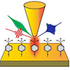
| (a) Schematic illustration of tip-enhanced VSFG spectroscopy. Tunable mid-IR- and near-IR-pulsed lasers are focused into the nanojunction between the STM tip and sample substrate, enabling the detection of VSFG signals from a very limited number of molecules located within the nanogap. (b) Observed VSFG spectra with fitted curves shown in black. (c) Extracted imaginary component of the vibrational resonant contribution of the second-order susceptibility ( Im[χR(2)(ωIR)]). Negative values indicate that the terminal methyl groups are oriented with hydrogen atoms directed away from the substrate. (Image: Atsunori Sakurai)
|
|
By precisely focusing both mid-IR and near-IR laser pulses into the narrow gap between the STM tip and the surface, the researchers were able to generate and detect sum-frequency signals from molecules directly underneath the tip. When the distance between the tip and the surface was large—around 50 nanometers—no signal was observed. But when the tip moved into the tunneling range, where the distance shrinks to less than a nanometer, a strong signal appeared. This confirmed that the technique depends on near-field effects to enhance the signal.
|
|
The researchers recorded eight different spectra by tuning the mid-IR wavelength. From these, they identified three distinct vibrational features that matched known signatures of terminal methyl groups on the molecules: symmetric stretching, a Fermi resonance between bending and stretching modes, and asymmetric stretching. Crucially, when the tip was more than 1 nanometer from the surface, the signal disappeared, showing that the detected VSFG response came from a region less than 1 nanometer in size.
|
|
This extremely tight confinement of light and signal was made possible by two interacting effects. First, the “antenna effect” concentrated IR light at the very tip of the probe. Second, the nanogap between the tip and the surface acted as a plasmonic cavity that enhanced the emission of the visible VSFG signal. Together, these effects enabled the detection of molecular vibrations from just a few molecules.
|
|
Additional analysis of the VSFG signal revealed a negative value in the imaginary part of the second-order nonlinear susceptibility, a parameter related to how the material interacts with the incoming light. This finding indicated that the methyl groups were oriented with their hydrogen atoms pointing away from the surface. In other words, the system could not only detect a small number of molecules but also determine how they were aligned.
|
|
In summary, this work demonstrates that it is possible to detect vibrational signals from molecules confined within a nanoscale region smaller than 1 nanometer. The approach provides both high spatial resolution and the ability to extract molecular orientation. This capability opens new possibilities for single-molecule spectroscopy and nanoscale molecular imaging, which could be valuable for studying catalytic surfaces and for designing more efficient catalytic materials.
|