Magnetic microrobots carry medicine through blood and release it only where needed, while being tracked inside the body using combined light and sound imaging.
(Nanowerk Spotlight) When a cancer drug enters the bloodstream, it cannot tell healthy cells from malignant ones. It travels wherever blood flows, spreading toxicity as it goes. The same is true for most medications. They move without direction, affecting far more tissue than the disease they are meant to treat. Precision therapy, where a drug reaches only its intended target, has remained a goal of modern medicine but not yet a reality.
Blood itself is the obstacle. It moves quickly, shifts direction, and passes through vessels so small that even bacteria struggle to enter. Magnetic particles can be moved by external fields, but only across short distances. Light can reveal their position, yet it fades inside living tissue. To create a drug carrier that can move, release, and be tracked inside the body, researchers needed new materials and new ways to see them.
The result is a microrobot that can carry medicine, travel through flowing fluid, release it in response to specific cues, and be tracked deep within tissue in real time. It functions as both vehicle and beacon.
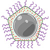
Schematic illustrations of a multifunctional magnetic microrobot platform (MMP) for targeted drug delivery applications. A) (i) Representation of a single engineered nanoparticle with a core-shell structure, featuring a hard magnetic FePt core and drug-loaded shell layer. (ii) Drugs are loaded into the shell (ZIF-8), (iii) with a spacious cavity in the center of the 3D structure of ZIF-8 for drug encapsulation. B) Scanning electron microscope (SEM) images of mPNs and the transmission electron microscope (TEM) image of a single mPN (scale bar: 200 nm (i) and 100 nm (ii)). C) (i) A weak magnetic field with the Girl with a Pearl Earring pattern. (ii) MMP demonstrates rapid responsiveness to small patterned, attractive magnetic field articles, exemplified by their interaction with the famous painting of “Girl with a Pearl Earring” pattern magnetic field. (scale bar: 1 cm). D) Illustration depicting the MMP (i) reconfigurable swarm behaviors, (ii) upstream movement, (iii) controlled drug delivery by PH and RF, and 3D real-time imaging with multi-spectral optoacoustic tomography (MSOT). (Image: Reprinted from DOI:10.1002/adma.202511870, CC BY) (click on image to enlarge)
The design begins with a hybrid nanoparticle. At its center is a magnetic core made of iron and platinum, written as FePt. Around it sits a porous shell formed from a metal–organic framework (MOF) called ZIF-8, short for zeolitic imidazolate framework 8. The FePt core provides permanent magnetization that allows stable steering. The ZIF-8 shell acts as a reservoir for drug molecules and breaks down in acidic environments such as those near tumors or inflamed tissue. Together they create a carrier that can be guided and triggered with precision.
Each microrobot measures about 200 nanometers across, small enough to move through narrow vessels but large enough to hold a meaningful dose. A coating of polyethylene glycol, known as PEG, reduces unwanted contact with proteins and shields the particles from immune recognition. Microscopy confirms the intended core shell structure. Magnetic testing reports a remanent magnetization of 24.70 electromagnetic units per gram and a coercivity of 153.68 millitesla. These values show that the particles maintain strong magnetization even after the external field is removed, which supports stable actuation.
The ZIF-8 shell offers ample space for drug molecules. Using the chemotherapy compound doxorubicin as a model, the particles achieved a loading efficiency of 93.9 percent after nine hours. When placed in acidic solution, drug release increased sharply, reaching almost complete release at pH 5.5. This mirrors the environment of tumor tissue.
The researchers also added external control. By applying radiofrequency energy at 338 kilohertz, they warmed the suspension to about 57 degrees Celsius in half an hour. The added heat accelerated release by promoting breakdown of the shell. These results show that release can be tuned by both local chemistry and external energy.
Motion control relied on collective behavior. When exposed to rotating or oscillating magnetic fields, many particles assembled into swarms that moved as single units. The fields could shape the swarms into chains, vortices, or diffuse clouds, each suited to a different task.
In test channels that simulated blood flow, the swarms traveled upstream against speeds of 70 millimeters per second and moved about 20 micrometers per second near vessel walls. In a branching channel, magnetic guidance directed 90.5 percent of the swarm into the chosen branch. Once at the target, a change in the magnetic pattern dispersed the swarm, preventing blockage in narrow paths.
Tests on living cells showed limited toxicity. Neuronal cells exposed to 10 micrograms per milliliter of particles retained about 87 percent viability after 48 hours. Immune assays recorded a short rise in inflammatory markers that subsided within two days.
A model of the blood brain barrier built from human brain endothelial cells showed that magnetic actuation increased particle transport from 20.14 percent to 78.9 percent within six hours. In co-culture experiments, doxorubicin loaded particles guided toward cancer cells achieved a level of cell death similar to that of free doxorubicin, while unloaded particles remained largely harmless.
Tracking the microrobots used a method called multispectral optoacoustic tomography, or MSOT. Short laser pulses illuminate the tissue, and absorbed light causes a rapid temperature rise that produces ultrasound waves. The returning sound is used to reconstruct three dimensional images with optical contrast and ultrasonic depth. The FePt ZIF-8 particles absorb strongly at about 920 nanometers, a wavelength where blood absorption is weak. This gives clear contrast deep inside tissue.
In pig brain samples the particles remained visible through seven centimeters of depth. Applying a magnetic field to concentrate them increased the signal and made real time tracking possible.
The particles also act as contrast agents for magnetic resonance imaging. Their presence shortens the T2 relaxation time in proportion to concentration, allowing imaging by standard MRI scanners. The combination of optoacoustic and magnetic contrast provides both live tracking and static verification.
Experiments in living mice showed how these elements work together. After injection into the bloodstream, the microrobots circulated through major vessels and were imaged continuously with MSOT. When a rotating magnetic field was applied, the swarms moved upstream through small vessels.
A computational technique called localization optoacoustic tomography processed the image sequence to identify moving points and sharpen vascular detail. In the brain, dynamic signals from the microrobots revealed arteries and veins as narrow as 22 micrometers and as deep as 1.5 millimeters, visible even through intact scalp and skull. Magnetic fields could trap and release the swarms at specific sites, demonstrating remote control within a living system.
The platform links drug loading, motion control, and imaging in a single framework. It shows that one type of particle can carry medicine, move with precision, and be seen as it travels. The study still faces practical limits. It does not yet prove that drugs can cross an intact blood brain barrier in living animals.
Applying the method to human tissue will require stronger magnetic fields and improved light delivery to overcome scattering. Long term safety must also be confirmed, since FePt fragments could persist in organs or pass through the kidneys depending on their size.
Even with these open questions, this work demonstrates a clear technical model for precise drug delivery. The FePt core provides magnetic strength. The ZIF 8 shell stores and releases medication in response to internal or external cues. PEG coating reduces immune interference. Swarm control enables motion in complex flow. Optoacoustic imaging and MRI make the process visible. The microrobots act not as passive carriers but as controllable agents capable of transport, release, and tracking in real time.
For authors and communications departmentsclick to open
Lay summary
Prefilled posts
ORCID information
Erdost Yildiz (Max Planck Institute for Intelligent Systems)
, 0000-0001-8086-3524 corresponding author
Metin Sitti (Max Planck Institute for Intelligent Systems)
, 0000-0001-8249-3854 corresponding author
Nanowerk Newsletter
Get our Nanotechnology Spotlight updates to your inbox!
Thank you!
You have successfully joined our subscriber list.
Become a Spotlight guest author! Join our large and growing group of guest contributors. Have you just published a scientific paper or have other exciting developments to share with the nanotechnology community? Here is how to publish on nanowerk.com.