Light responsive nanomotors dissolve blood clots and repair vessel tissue by combining catalytic activity, targeted motion, and gentle heating for coordinated thrombolysis and endothelial regeneration.
(Nanowerk Spotlight) When a blood vessel is injured, the body forms a clot to seal the break and prevent bleeding. Platelets gather at the site and become embedded in a mesh of fibrin strands. This plug normally dissolves once healing is complete. When it does not, the clot can block circulation and deprive tissues of oxygen.
Medicines that dissolve fibrin can reopen flow, but they leave the vessel lining fragile and inflamed. That lining, called the endothelium, may continue releasing reactive oxygen species, unstable oxygen-based molecules that damage cells and attract more platelets. The result is a feedback loop that favors renewed blockage.
Scientists have tried to solve this with targeted nanoparticles, light-responsive materials, and catalytic coatings that remove reactive oxygen species. Each method offers part of the answer. Platelet membranes can guide particles to injured vessels but cannot penetrate the dense fibrin network. Near infrared light can gently warm the clot and soften its fibers but may cause local overheating if not controlled. Catalytic membranes can reduce oxidative stress but cannot direct themselves to the right location. No single system has combined all of these properties in a way that allows precise clot removal and restoration of vessel health.
A study published in Advanced Materials (“NIR‐Driven Nanomotors Integrating With Platelet‐Thylakoid Hybrid Membranes for Synchronized Thrombolysis and Vascular Remodeling”) reports a design that brings these functions together. It describes nanoscale particles that move on their own inside clots and respond to near infrared light to enhance their motion and reactivity. The goal is to dissolve fibrin while calming inflammation and encouraging repair of the vessel lining. The researchers tested the particles in cell cultures, artificial clots, zebrafish, and a mouse artery injury model.
The nanomotor has a core made of mesoporous polydopamine that contains strontium ions and a shell formed from a blend of platelet and thylakoid membranes. Mesoporous means the core has nanosized pores that create a large surface area for reactions. Polydopamine is a synthetic version of the adhesive polymer found in mussel shells that efficiently converts near infrared light into mild heat. The platelet membrane directs the particle to injured endothelium, the thylakoid membrane contains enzymes that act like catalase and convert hydrogen peroxide into oxygen and water.
Hydrogen peroxide is abundant inside a clot, so this reaction provides both propulsion and detoxification. The oxygen that forms propels the particle forward in tiny bursts while reducing oxidative damage. The researchers refer to the construct as PSr at PT, where PSr is the polydopamine and strontium core and PT is the platelet and thylakoid coating.
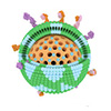
Designing pathogenesis-adaptive polydopamine nanomotors (PSr@PT NPs) for synchronized thrombolysis and vascular microenvironment remodeling. A) Schematic illustration of the PSr@PT NPs synthesis process, and mechanisms of inhibition of thrombosis and thrombolysis by PSr@PT NPs. B) PSr@PT NPs are capable of achieving platelet membrane-mediated targeting to injured endothelium, generating gas bubbles to facilitate the self-propelled motion of the nanoparticles. They alleviate oxidative stress, photothermally enhance the penetration and dissolution of clots, and further promote the repair of the vascular microenvironment. (Image: Reprinted with permission by Wiley-VCH Verlag) (click on image to enlarge)
The concept is to use the chemical environment of the clot as fuel. High hydrogen peroxide triggers movement and detoxification. Near infrared light gently raises the temperature to around forty degrees Celsius, loosening fibrin fibers and helping the nanomotors move deeper into the clot. Inside this oxidative environment, the core releases strontium ions, which act as biochemical cues that promote endothelial cell growth and tissue repair.
The result is a synchronized process of clot removal and vessel recovery.
Microscopy shows that the particles are about one hundred ninety five nanometers wide with a clear core and shell. Element mapping places strontium inside the core and phosphorus, a component of membranes, on the surface. Protein analysis identifies markers typical of platelets and thylakoids, showing that both types of membrane remain intact. The particles remain stable under conditions similar to those found in blood.
Tests of catalytic and thermal properties confirm that the coated particles perform as designed. Both bare polydopamine cores and the coated nanomotors neutralize free radicals, but only the coated version decomposes hydrogen peroxide strongly and generates sustained oxygen bubbles. When illuminated with near infrared light at one watt per square centimeter, the suspension warms from twenty two to about forty one degrees Celsius in ten minutes.
The photothermal conversion efficiency is about 34 percent and remains constant during repeated heating and cooling cycles. This mild temperature rise is sufficient to soften fibrin without harming surrounding tissue in the reported tests.
Motion tracking links chemical reactions to propulsion. In pure water the nanomotors move randomly. In a solution containing hydrogen peroxide they move along curved paths and travel farther. Near infrared light alone increases movement slightly due to thermal gradients. The combination of hydrogen peroxide and light produces the strongest motion.
The same pattern appears in serum, indicating that proteins do not inhibit mobility. When placed in a gradient of hydrogen peroxide, the particles migrate toward the higher concentration, suggesting that they could naturally move into the oxidative core of a clot.
Cell experiments examine targeting, protection, and anti inflammatory effects. Inflamed endothelial cells take up far more coated particles than healthy ones, and more than uncoated cores. Under conditions of oxidative stress and low oxygen, cells treated with the coated particles remain viable and show reduced reactive oxygen species.
Imaging shows restored mitochondrial potential, which signals healthy energy balance. Levels of inflammatory molecules such as tumor necrosis factor alpha and interleukin six fall sharply. The treated cells release less von Willebrand factor, a protein that attracts platelets. As a result, platelets adhere much less to the treated cell surfaces. These outcomes indicate that the nanomotors not only dissolve fibrin but also calm the environment that sustains thrombosis.
The same particles stimulate repair. In standard assays, treated endothelial cells form more tube like networks, and levels of vascular endothelial growth factor increase. In a chicken embryo membrane test, which measures new vessel formation, the formulation promotes more blood vessels, consistent with strontium’s known role in cell signaling and tissue regeneration.
Proteomic analysis connects these effects to broader molecular pathways. Endothelial cells under stress show different expression of one hundred forty four proteins after treatment. Many are linked to clot formation, breakdown, and endothelial function. Three signaling pathways are affected. JAK STAT, which regulates inflammation, PI3K Akt, which controls cell survival, and platelet activation signaling all show reduced activity. Levels of fibronectin, a structural protein that supports repair, increase.
In artificial clots, near infrared light helps the nanomotors penetrate more deeply. The combination of light and nanomotors produces faster clot shrinkage and higher levels of fibrin breakdown products. The fibrin mesh becomes looser and more porous. Tests on red blood cells show no significant hemolysis within the studied concentration range, indicating good compatibility.
Animal studies evaluate both prevention and treatment. In zebrafish, a chemical trigger creates thrombosis. Because the fish are small and move constantly, light cannot be directed precisely, so the tests measure the chemical activity alone. Whether the particles are added to water or injected, they restore blood flow and lower reactive oxygen signals in the region where clots form. The clots appear smaller in stained images of treated fish.
In mice, the team uses a ferric chloride injury to block the carotid artery. In preventive experiments, animals receive nanomotors before injury followed by local near infrared exposure. Laser imaging shows that blood flow stays close to baseline levels, while untreated controls show near total blockage. Tissue analysis reveals smaller clots and fewer markers of oxidative DNA damage and hypoxia. The vessel shows higher levels of proteins related to barrier integrity and smooth muscle structure, suggesting improved healing. Blood chemistry and tissue sections show no signs of toxicity.
A therapeutic protocol tests existing clots. Fluorescent imaging confirms that the platelet coating guides the nanomotors directly to the thrombus. When exposed to near infrared light at one watt per square centimeter for ten minutes, blood flow returns to nearly normal within a day. Markers of oxidative stress and platelet accumulation fall, while proteins linked to vessel stability and nitric oxide production rise. Repeated light exposure causes no visible or microscopic skin damage under these conditions.
Across experiments the findings follow a consistent pattern. The platelet coating ensures the particles reach the damaged site. The thylakoid enzymes convert hydrogen peroxide into oxygen, reducing oxidative stress and producing motion. Polydopamine provides controlled heating that softens fibrin and improves penetration. Strontium ions support the regrowth of endothelial tissue. Together these processes allow one system to both dissolve the clot and repair the surrounding vessel.
This study presents a method that links clot removal with recovery of the vessel environment. It demonstrates coordinated effects in cell cultures and animal models and outlines a mechanism for precise control through light. The work stops short of pharmacological testing or long term safety analysis but establishes a clear proof of concept. A light guided nanomotor that acts on both the clot and the vessel may offer a path toward treatments that restore blood flow while supporting tissue repair.
For authors and communications departmentsclick to open
Lay summary
Prefilled posts
Nanowerk Newsletter
Get our Nanotechnology Spotlight updates to your inbox!
Thank you!
You have successfully joined our subscriber list.
Become a Spotlight guest author! Join our large and growing group of guest contributors. Have you just published a scientific paper or have other exciting developments to share with the nanotechnology community? Here is how to publish on nanowerk.com.