Erasable electric fields written by laser light onto crystal surfaces can trap, align, and release living cells without electrodes or physical contact.
(Nanowerk Spotlight) All human cells possess electrical properties. Every cell maintains a voltage difference across its membrane, produced by the movement of charged ions like sodium, potassium, and calcium. Many cell types can actively respond to electric fields, though not all are specialized to sense them. In skin, for example, ion transport creates a measurable voltage difference between inner and outer tissue layers.
When a wound breaks the skin, that voltage difference collapses locally, producing an electric field that radiates outward from the injury site. Nearby cells, including fibroblasts and immune cells, detect this field and migrate toward the wound to close it. Similar endogenous electric signals guide nerve fibers toward their targets during development and establish the structural organization that distinguishes one side of a tissue from another.
This natural electrical sensitivity has attracted bioengineers for a practical reason: if certain cells already follow electric field cues, then artificially generated fields might direct those cells on command. Controlling where cells move, how they attach, and what shape they adopt would open direct paths to growing replacement tissues, guiding nerve repair, and studying how diseases exploit cell migration.
The standard approach has been to build electrode arrays into cell culture substrates, applying voltages to push and pull cells into desired positions. These systems have produced useful results, but they share a fundamental limitation. Electrodes are fabricated through multi-step lithographic processes that embed conductive materials into biocompatible surfaces, and once built, the patterns cannot change. Altering the stimulation geometry means redesigning and rebuilding the entire device. External wiring and power supplies add further complexity.
Ferroelectric crystals, materials whose internal electric charges can be manipulated, offered a partial way forward. Lithium niobate, one such crystal, showed early promise. Studies demonstrated that its natural surface charge could influence how fibroblast cells adhere and spread.
Other work showed that periodically reversing the crystal’s internal polarization could partially guide the growth of cortical nerve fibers along domain boundaries. Pyroelectric patterning of polymer sheets in contact with lithium niobate achieved selective adhesion of neuroblastoma cells. And iron-doped lithium niobate proved capable of trapping and orienting bacteria through its photovoltaic response to light.
Each result confirmed that ferroelectric surfaces could interact meaningfully with biological material. None achieved real-time, reversible control over living cells that were attached and growing on the surface.
A study published in Advanced Functional Materials (“An All‐Optical Driven Bio‐Photovoltaic Interface for Active Control of Live Cells”) addresses that limitation. A research team based primarily at the Institute of Applied Sciences and Intelligent Systems of the Italian National Research Council developed what it terms a bio-photovoltaic interface.
The platform uses iron-doped lithium niobate crystals whose surfaces generate tunable electric fields under laser illumination. These fields trap fibroblast cells, direct their alignment, deform their nuclei, and, when the light erases the stored charge pattern, release them. No electrodes, wiring, or permanent surface modification are involved.
Fabrication of the bio-photovoltaic interface. (A) laser induced optical activation of the photovoltaic field at the air-crystal interface and formation of the volume phase grating; (B) comparison between laser-light profile and charge distribution by Comsol Multiphysics simulation for the evaluation of charges location and field strength; (C) sketch of the experimental procedure for cell seeding made of Fe:LN cleaning and UV sterilization, and subsequent cell plating. The sketch shows how the cells are first trapped and, after 24 hours, present a pronounced polarization along the c-axis of the crystal. (Image: Reproduced from DOI:10.1002/adfm.202518941, CC BY) (click on image to enlarge)
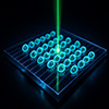
The physical mechanism centers on the photovoltaic effect in iron-doped lithium niobate. When the crystal absorbs light at an appropriate wavelength, photons drive charge redistribution within the material, producing internal electric fields that can reach 10⁶ V/cm. These internal fields generate a weaker electric field just above the crystal surface that fades rapidly with distance. By projecting an argon laser beam at 514 nm through a patterned optical mask, the researchers inscribed periodic stripe patterns into the crystal at spacings of 25 µm and 50 µm. The same process modulates the crystal’s refractive index, creating an internal optical grating that confirms the pattern has been successfully written.
The team seeded NIH-3T3 mouse fibroblast cells onto four substrates: iron-doped lithium niobate patterned at 50 µm (PV-50), the same material patterned at 25 µm (PV-25), unpatterned lithium niobate, and plain glass. The team plated cells at identical density and incubated them for 24 hours. On both patterned substrates, approximately 80% of cells oriented perpendicular to the grating stripes, aligning their long axes along the electric field gradient. Cells on the two control surfaces attached without any preferred direction. Statistical testing confirmed highly significant differences between patterned and control groups (adjusted p < 0.0001).
The electric fields also reshaped cell nuclei. Fluorescence imaging showed that nuclei on PV-50 had a mean elongation index of 0.5 (where 0 represents a circle and 1 a maximally stretched shape), while PV-25 nuclei averaged 0.4. Both control surfaces produced nearly circular nuclei at 0.1. Statistical analysis confirmed these differences were highly significant (p < 0.001) and attributable to the photovoltaic patterning rather than the substrate material itself.
Confocal microscopy revealed the structural basis for these changes. On patterned substrates, actin filaments, the protein fibers that form a cell’s internal scaffolding and transmit mechanical forces, reorganized along the electric field direction. This cytoskeletal tension propagated to the nucleus through the LINC complex, a molecular bridge connecting the actin network to the nuclear envelope.
Cells on PV-50 developed the strongest adhesion and nuclear deformation. The wider 50 µm spacing better matched natural cell dimensions, allowing each cell to spread fully within a single confinement zone and maximize mechanical signaling. The tighter 25 µm period restricted spreading and fragmented electric cues, producing weaker alignment despite theoretically stronger spatial confinement.
Digital holographic microscopy added a real-time dimension to these observations. This label-free technique measures the optical path delay through transparent specimens, producing quantitative maps of cell shape and position without any staining. Living cells monitored for 20 hours on PV-50 remained confined to specific zones of the grating. Their nuclei stayed in place while protrusions extended and retracted across adjacent regions. Cells elongated when their nuclei occupied one zone and became rounder when drifting toward the neighboring zone, which acted as a repellent barrier.
Erasing the pattern changed cell behavior dramatically. When the laser redistributed stored charges back to a uniform state, confined cells regained mobility. A tracked cell covered three times the distance it had managed under confinement. In separate experiments using discrete dashed-line barriers, fewer than 10% of cells crossed an active barrier at low cell densities.
After optical erasure, cells migrated freely through the same region. A live/dead viability assay confirmed the platform’s safety: cell survival ranged from 91.7% on PV-25 to 93% on glass, with no meaningful difference between patterned and control substrates.
The platform requires no lithographic fabrication, no electrode wiring, and no external power supply. Patterns can be written, erased, and rewritten on the same crystal using structured light alone, and the holographic microscope simultaneously tracks both the electric field pattern and the cellular response.
The researchers note that current fabrication uses laser powers too high to apply while cells are present, so pattern writing must precede cell seeding. Only the erasure step operates safely alongside living cultures. Future work aims to develop conditions for inscribing patterns directly in the presence of cells.
Applications could span tissue engineering, where directed cell alignment is needed to build functional constructs, and neuroscience, where a reconfigurable stimulation surface could replace the fixed electrode arrays now used to study neural signaling.
For authors and communications departmentsclick to open
Lay summary
Prefilled posts
ORCID information
Lisa Miccio (Institute of Applied Sciences and Intelligent Systems of CNR (CNR-ISASI))
, 0000-0001-9427-881X corresponding author, first author
Nanowerk Newsletter
Get our Nanotechnology Spotlight updates to your inbox!
Thank you!
You have successfully joined our subscriber list.
Become a Spotlight guest author! Join our large and growing group of guest contributors. Have you just published a scientific paper or have other exciting developments to share with the nanotechnology community? Here is how to publish on nanowerk.com.