Graphene oxide selectively destroys bacteria by bonding with a membrane lipid absent from human cells, killing microbes while leaving healthy tissue unharmed.
(Nanowerk Spotlight) Most antibacterial nanomaterials damage human cells along with bacteria. Silver nanoparticles kill bacteria through ion release but cause oxidative stress in healthy tissue. Copper nanoparticles share the same limitation. Carbon nanotubes puncture bacterial membranes with their needle-like shape, yet that geometry injures mammalian cells equally. Among antibacterial nanomaterials, potency against bacteria and safety for human tissue have not been achievable simultaneously.
Graphene oxide, an oxidized form of graphene bearing abundant oxygen functional groups on its surface, defies this constraint. A 2024 clinical study published in Nature Nanotechnology found that human subjects who inhaled small graphene oxide sheets experienced no adverse pulmonary, cardiovascular, or inflammatory responses. Yet in laboratory settings, the same material suppresses bacterial growth at rates exceeding 99%. These two properties have coexisted without a molecular explanation. Proposed mechanisms, including membrane cutting, oxidative damage, and lipid extraction, have derived primarily from computational simulations and do not account for the selectivity that spares mammalian cells.
A study published in Advanced Functional Materials (“Biocompatible but Antibacterial Mechanism of Graphene Oxide for Sustainable Antibiotics”) now provides direct experimental evidence for that selectivity. The research team traced graphene oxide’s antibacterial action to a specific hydrogen-bonding interaction with a phospholipid found exclusively in bacterial membranes, a molecule absent from mammalian cells. This finding reframes the apparent contradiction between biocompatibility and antibacterial efficacy as two expressions of a single selective mechanism.
Schematic illustration of the selective antibacterial mechanism of GO nanosheets and their chemical structures. (Image: Reproduced from DOI:10.1002/adfm.74695, CC BY) (click on image to enlarge)
The team prepared graphene oxide nanosheets with systematically varied chemical properties, adjusting flake size, oxygen content, and nitrogen doping. Unmodified graphene oxide, carrying its full load of oxygen functional groups, suppressed more than 99% of E. coli growth. Thermal reduction progressively stripped away oxygen and weakened the effect: moderately reduced sheets achieved 68% suppression, and fully reduced sheets only 57%. Nitrogen doping dropped efficacy to 60%. Oxygen content proved to be the decisive variable.
To understand why, the team built artificial vesicles mimicking bacterial membranes. Bacterial membranes contain a phospholipid called POPG that constitutes more than 15% of their lipid content but does not appear in mammalian cell membranes. A second phospholipid, POPE, is common to both. The researchers constructed vesicles from each lipid separately, then exposed them to graphene oxide.
Cryogenic electron microscopy showed that graphene oxide selectively destroyed POPG-containing membranes, producing punctured and collapsed structures. Pure POPE membranes remained intact. Infrared spectroscopy confirmed that POPG vesicles absorbed large quantities of graphene oxide flakes through specific chemical binding, while POPE vesicles did not.
Nuclear magnetic resonance spectroscopy pinpointed the bonding mechanism. POPG carries a diol group, two hydroxyl groups on its glycerol headgroup, that forms hydrogen bonds with the oxygen functional groups on graphene oxide’s surface. The team verified this interaction using model compounds representing graphene oxide’s typical functional groups, including alcohols, epoxides, and carboxylic acids, all of which produced the same characteristic spectral shifts when mixed with POPG.
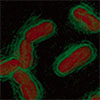
Three-dimensional holotomography, a technique that images living cells without staining, captured the consequences in real time. E. coli cells exposed to graphene oxide gradually lost their rod shape, swelled, and ruptured, spilling their contents. Scanning electron microscopy confirmed surface wrinkling followed by structural collapse.
The researchers incorporated graphene oxide into nylon nanofibers at 1 and 2 wt% using electrospinning, producing fibers roughly 200 nm in diameter. These composite fibers killed bacteria effectively and retained their activity after thorough washing, confirming stable integration of graphene oxide within the polymer. Cytotoxicity tests on mammalian fibroblast cells showed minimal harm over 72 hours.
The choice of polymer mattered: polyvinyl alcohol fibers lost their antibacterial function because the polymer’s own hydroxyl groups competed for hydrogen bonding with graphene oxide, blocking its access to bacterial membranes. Nylon, by contrast, relies on interchain hydrogen bonding between its own amide groups, leaving graphene oxide’s surface functional groups available to interact with bacteria.
In mice with infected full-thickness skin wounds, graphene oxide applied as film, nanofiber, or powder substantially reduced E. coli counts within 7 days. Powder eliminated detectable colonies by day 3. All formats accelerated wound closure and produced minimal inflammation in tissue analysis.
The team then tested the materials in pigs, whose skin closely resembles human skin. Nanofibers achieved greater than 99.9% bacterial reduction within 9 days, outperforming the other formats because their flexibility maintained better tissue contact and prevented material loss from the wound. Histological examination confirmed minimal bleeding and inflammation.
Graphene oxide also suppressed K. pneumoniae, strains of both E. coli and K. pneumoniae carrying the NDM-1 resistance gene, and methicillin-sensitive and methicillin-resistant S. aureus. The antibacterial mechanism for Gram-positive species such as S. aureus may differ from the POPG-targeting pathway observed in Gram-negative bacteria, however, given the thick peptidoglycan layer surrounding their membranes.
Because POPG is a conserved and essential structural component of bacterial membranes, bacteria cannot easily shed it without compromising their own survival. This makes the graphene oxide mechanism fundamentally different from conventional antibiotics, which target specific enzymatic pathways that bacteria can mutate around. The nanofibers also retained their antibacterial function after repeated washing, raising the prospect of reusable wound dressings and protective medical textiles.
A material that exploits a structural feature bacteria cannot afford to lose, rather than a biochemical target they can evolve to protect, offers a durable complement to conventional antibiotics in settings where drug resistance is accelerating.
For authors and communications departmentsclick to open
Lay summary
Prefilled posts
ORCID information
Sang Ouk Kim (Korea Advanced Institute of Science and Technology (KAIST))
, 0000-0003-1513-6042 corresponding author
Nanowerk Newsletter
Get our Nanotechnology Spotlight updates to your inbox!
Thank you!
You have successfully joined our subscriber list.
Become a Spotlight guest author! Join our large and growing group of guest contributors. Have you just published a scientific paper or have other exciting developments to share with the nanotechnology community? Here is how to publish on nanowerk.com.