Scientists create a biohybrid by combining E. coli with a metal-organic framework, enabling charge storage through microbial electron transfer.
(Nanowerk Spotlight) Harnessing biological systems for electronic functionality offers the potential to build devices that are compact, self-sustaining, and responsive to living environments. Microorganisms, with their rich metabolic activity and adaptive biochemistry, are especially attractive as components of bioelectronic systems.
However, to integrate living microbes into functional devices, researchers need a reliable way to capture and control the tiny electrical signals that their cells generate. These signals originate from electron movement during metabolism and could, in principle, be converted into measurable current or stored charge.
If this electron flow can be extracted in a stable and controlled fashion, it becomes possible to design microbial sensors, biological batteries, or programmable biocapacitors—devices that store and discharge energy based on live cell activity. Such systems could be used in environmental monitoring, biomedical diagnostics, or energy-harvesting applications where conventional electronics cannot function as efficiently or sustainably. Yet this approach has remained largely confined to a few specialized bacterial species that naturally expel electrons to their surroundings.
The majority of microbes, including common strains like Escherichia coli, do not possess this capacity. Without a natural pathway to transfer electrons beyond the cell membrane, these organisms are not compatible with existing bioelectronic designs. This limitation has prompted researchers to explore new materials and interfaces that might coax even non-electrogenic cells into releasing electrical charge externally.
Among the most promising candidates are metal-organic frameworks (MOFs), synthetic porous nanomaterials that can be engineered to interact with biological systems at the molecular level.
The bacterium, when physically coupled with this MOF, becomes part of a biohybrid structure that enables charge movement from the bacterial membrane to an electrode. This results in a measurable and stable electrical signal, allowing the system to function as a live-cell biocapacitor.
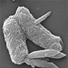
Schematic illustration of the chemistry for the preparation ofMIL-88B(Fe)-MOF and the formation of the biohybrids, E.coli/MIL-88B(Fe)-MOFs. (Image: reprinted from DOI:10.1002/smll.202411472, CC BY)
The researchers created the biohybrid by incubating E. coli with MIL-88B(Fe) particles. This MOF contains trimeric iron(III) nodes, which serve as redox-active centers. High-resolution imaging confirmed that the MOF particles bind directly to the bacterial surface. Spectroscopic data showed a shift in the oxidation state of the iron ions within the MOF, consistent with electron uptake from the bacteria. The shift from Fe(III) to Fe(II) indicates that the MOF accepts electrons and participates in a redox reaction with the bacterium.
Control experiments established that neither E. coli nor MIL-88B(Fe) alone produced significant electron transfer to an electrode. Only the hybrid configuration generated measurable electrical signals, demonstrating that the interface between the bacterium and the MOF is essential for the observed behavior.
Conductive atomic force microscopy and Kelvin probe force microscopy showed that when placed on an electron-deficient polymer substrate, the biohybrid produced higher surface potential gradients than either of its components alone. This provided direct evidence of electron movement from the bacteria, through the MOF, to the substrate.
The team then examined the system’s ability to store electrical charge. Cyclic voltammetry measurements showed that the hybrid exhibits a combination of two types of capacitance. The first, known as electrical double-layer capacitance, arises from surface charge accumulation. The second, pseudocapacitance, results from reversible redox reactions involving the MOF’s metal centers. The hybrid’s voltammetry curves showed a distinct reduction peak around −0.053 V, corresponding to the conversion of Fe(III) to Fe(II).
The hybrid demonstrated a total specific capacitance of 431 F/g, as calculated using the Trasatti method, which separates surface-based and redox-based charge contributions. Of this total, 177 F/g came from surface charge, while 254 F/g was attributed to pseudocapacitance—nearly double the pseudocapacitive contribution of the MOF alone (142 F/g).
In comparison, E. coli alone produced a total capacitance of only 195 F/g at low scan rates and was electrochemically inactive at higher scan rates. The fivefold enhancement over E. coli alone confirms that the MOF was responsible for initiating and sustaining electron transfer.
Further analysis showed that this increase in performance did not compromise bacterial viability. After electrochemical testing, colony-forming assays demonstrated that E. coli cells remained alive and capable of growth. This is an essential requirement for live-cell biocapacitor applications, which rely on ongoing biological activity.
Additional charge–discharge tests showed that the hybrid retained its charge over time and could release it in a controlled way. Galvanostatic discharge curves revealed longer discharge times for the hybrid compared to either component. Electrochemical impedance spectroscopy further confirmed that the hybrid interface had lower charge transfer resistance than E. coli or the MOF alone. The hybrid’s Nyquist plots exhibited steeper slopes and smaller semicircles, signatures of improved electron transport and reduced internal resistance.
The MOF’s role as a mediator is essential to this function. Its redox-active metal centers can undergo partial reduction when in contact with electrons released from the bacterium. This provides a conductive pathway for electrons to move toward the electrode. In this system, MIL-88B(Fe) acts as a synthetic bridge, capturing metabolic electrons that would otherwise remain confined within the cell and facilitating their flow to an external circuit. The result is a functioning biocapacitor that uses live E. coli as an electrochemical component.
The work demonstrates that microbial electron transfer can be induced without genetic modification or external mediators in solution. Instead, the material interface itself, specifically the MOF, performs the role of mediating and storing charge. This capability introduces a new class of bioelectronic devices that use viable, unmodified cells as active materials.
By enabling a controllable electron transfer process in E. coli, a species not previously considered suitable for such applications, the study outlines a general strategy for integrating other non-electrogenic microbes into electrochemical systems. The results also show that MOFs can be tailored not only for compatibility with biological systems but also to actively participate in signal transduction and energy storage.
This biohybrid system points to future devices that could operate in dynamic environments, such as implantable sensors or environmental monitors, where biological inputs need to be converted into readable electronic outputs. The integration of living cells into charge-storing systems may also allow for new forms of autonomous diagnostics or self-powered microdevices. As synthetic biology, nanomaterials, and electrochemistry continue to intersect, the concept demonstrated here may become foundational for the next generation of biologically integrated electronics.
Get our Nanotechnology Spotlight updates to your inbox!
Thank you!
You have successfully joined our subscriber list.
Become a Spotlight guest author! Join our large and growing group of guest contributors. Have you just published a scientific paper or have other exciting developments to share with the nanotechnology community? Here is how to publish on nanowerk.com.