Biodegradable microneedles act as binary fluorescent switches, encoding pH and glucose levels beneath the skin into scannable QR codes that require no calibration.
(Nanowerk Spotlight) Just beneath the skin’s surface lies a thin layer of fluid that mirrors much of the blood’s biochemistry. This extracellular fluid carries glucose, ions, and other small molecules in concentrations that track conditions from wound infection to diabetic crisis, and unlike blood, it can be reached without drawing from a vein. Microneedles, roughly a millimeter long, penetrate painlessly into the upper skin layers and make direct contact with this reservoir.
Microneedles were originally developed for transdermal drug delivery, but researchers have adapted them for biosensing by incorporating responsive materials into the needle tips. Early electrochemical versions wired each needle to a readout circuit that stayed attached to the skin. Colorimetric variants changed color in response to analytes such as glucose or pH, but subtle color shifts are hard to read through skin, and nonlinear calibration curves complicate clinical translation.
Fluorescence-based microneedles offered higher sensitivity and the possibility of wireless readout. Yet these too rely on measuring a continuously varying signal intensity. Differences in insertion depth, tissue scattering, probe diffusion, and ambient light all distort the analog readout, causing calibration drift and poor reproducibility from one patient to the next.
A study published in Advanced Materials (“Digital Microneedles for Multiplexed Transdermal Sensing via Fluorescent QR Codes”) now takes a fundamentally different approach. Instead of reading exact concentrations from graded fluorescence, the researchers engineered microneedles that behave like binary switches. Each needle contains a fluorescent probe tuned to switch between “off” and “on” at a single, predefined analyte concentration. The readout no longer depends on how bright a needle glows, only on whether it glows at all.
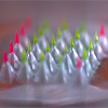
By arranging 25 such needles in a 5 × 5 grid, the team generated a two-dimensional fluorescent pattern that functions as a scannable QR code. Because every needle makes only a yes-or-no decision, the system sidesteps calibration entirely.
Conceptual framework contrasting analog vs. digital fluorescent microneedle sensing. (a,b) Schematic workflow: patch application and tip detachment after skin insertion (a), followed by optical readout by blue-light illumination producing a scannable QR code that dynamically reflects analyte concentrations (b). Digital microneedles in (b) exhibit sharp, nonlinear activation at defined thresholds, functioning as binary switches that eliminate calibration and reduce ambiguity. (c) Analog microneedles, in contrast to digital ones, generate graded fluorescence intensity as a function of analyte concentration, requiring calibration and remaining prone to variability from tissue and environmental conditions, leading to gain and offset errors. (d) Staggered thresholds across multiple microneedles generate binary fluorescent codes corresponding to analyte concentration ranges. (e) Arrangement of digital microneedles in 2D arrays produces fluorescent QR codes whose patterns dynamically change with analyte levels, enabling direct optical decoding of biochemical information. Green fluorescence indicates sensing microneedles, while red fluorescence marks reference microneedles for orientation and insertion confirmation. (Image: Reproduced from DOI:10.1002/adma.202518935, CC BY) (click on image to enlarge)
The microneedles adopt what the researchers call a mechanically optimized “baby-bottle” shape, designed to balance two competing demands: reliable skin penetration and controlled tip separation afterward. A narrow conical tip sits atop a wider pedestal, and a trapped air bubble separates the two. The bubble preserves rigidity under downward compression during insertion but weakens the joint under lateral shear, so a simple sideways thumb motion snaps the tips free from the backing patch. The tips stay embedded in the skin while the patch is peeled away.
All materials are biodegradable. Each tip uses a core-shell architecture: a rigid poly(lactic-co-glycolic acid) core provides structural support, while a thin polyvinyl alcohol shell houses the fluorescent probe. Upon contact with interstitial fluid, the shell swells into a hydrogel that lets small molecules diffuse inward to reach the probe.
Fabrication relies on a 3D-printed master mold replicated in a silicone negative. Sensing needles receive a polyvinyl alcohol solution doped with a green fluorescent probe, centrifuged into a thin shell lining the cavity walls before the polymer core is cast on top. Reference needles are filled with core polymer containing a red dye. A sugar-polymer solution forms the pedestal and backing, trapping the air bubble by surface tension. The finished patch is a circular disc roughly 1.5 cm across, with a maximum dimensional coefficient of variation of 4.3% across batches.
Can the needles penetrate skin and detach cleanly? Tests in porcine skin showed entry at approximately 0.15 N per needle, well below the force that would deform the tips. Detachment required only about 0.01 N of lateral force. Under realistic thumb-press application, penetration efficiency reached 98% and detachment efficiency 91%. Cytotoxicity testing with human dermal fibroblasts confirmed that the materials and loaded probes caused no detectable harm to cells.
What makes the system digital rather than analog is how each probe is tuned to act as a threshold switch. For pH sensing, the team used fluorescein at varying concentrations across the array. At low probe loading, a needle lights up only at high pH; at higher loading, it activates at lower pH. This creates a staircase of thresholds spanning pH 4.5 to 8.5 in 1 pH unit steps, a resolution relevant to wound care, where clinically meaningful pH shifts between healthy and chronically infected tissue are often on that scale.
For glucose, fluorescein boronic acid served as the probe, but the logic is inverted: fluorescence starts bright and drops sharply once glucose exceeds a set level. Thresholds were spaced at 2 mM intervals across a 1 to 10 mM range, yielding an uncertainty of ±1 mM.
That figure is comparable in magnitude to the accuracy criterion in international standards for self-monitoring blood glucose systems, suggesting the approach could be clinically actionable even at this early proof-of-concept stage. For both analytes, the contrast between on and off states was large enough that a simple brightness cutoff could reliably classify each needle.
To test simultaneous detection of both analytes in real skin, the researchers fabricated a combined QR code patch. It carried nine pH-sensing needles, nine glucose-sensing needles, and seven red-dye reference needles for orientation. Patches were inserted into porcine skin incubated in interstitial fluid at various pH and glucose combinations.
The fluorescent pattern stabilized about 30 minutes after insertion, consistent with the diffusion time through the polyvinyl alcohol shell and remained accurately decodable for approximately 2.5 hours beyond that settling period. Within this operating window, the digital output was reversible: alternating the surrounding analyte conditions produced reproducible QR code switching with the same 30-minute stabilization time.
A custom image-processing algorithm segmented each fluorescence image into a 5 × 5 grid, classified each needle as on or off, and applied majority-vote logic across triplicate needles for each threshold. Across 27 independent patches, classification accuracy reached 93% for pH and 85% for glucose. The triplicate design builds in fault tolerance: if one needle fails to insert properly, the remaining two still deliver a correct majority vote.
The operational window is limited by degradation of the polyvinyl alcohol matrix after a few hours, with the polymer core dissolving fully within 14 days. The quantization uncertainty of ±0.5 pH units and ±1 mM glucose is coarser than what analog sensors promise on paper, but it is a deliberate trade-off for robustness in a transdermal environment where optical variability makes analog readings unreliable.
By replacing analog intensity measurement with a binary decision at each needle, this platform reframes the engineering challenge: rather than fighting tissue variability with ever-more-complex calibration, it renders that variability irrelevant. The modular architecture offers a direct path to finer resolution through larger arrays with more densely spaced thresholds, and the concept extends to any analyte for which a threshold-responsive fluorescent probe exists.
Future work will need to extend the readable time window, sharpen probe switching characteristics, and validate performance in living animals. If those hurdles are cleared, a smartphone camera fitted with a small clip-on filter module could capture and decode these fluorescent QR codes at the point of care, turning a disposable skin patch into a multiplexed diagnostic tool for wound management and diabetes monitoring.
For authors and communications departmentsclick to open
Lay summary
Prefilled posts
Nanowerk Newsletter
Get our Nanotechnology Spotlight updates to your inbox!
Thank you!
You have successfully joined our subscriber list.
Become a Spotlight guest author! Join our large and growing group of guest contributors. Have you just published a scientific paper or have other exciting developments to share with the nanotechnology community? Here is how to publish on nanowerk.com.