A DNA hydrogel engineered from tetrahedral nanostructures accelerates burn wound healing by directing immune cell behavior, reducing inflammation, and supporting tissue regeneration through structural and biochemical interactions.
(Nanowerk Spotlight) Some burn wounds heal without complications. Others don’t. In more severe cases, the damage doesn’t end with the initial injury. It can trigger a persistent immune response that stalls healing, turning what should be a temporary wound into a chronic, inflamed lesion. The body’s natural repair process, normally a tightly regulated sequence of inflammation, tissue rebuilding, and regeneration, breaks down. Instead of resolving the damage, immune cells linger, releasing harmful molecules that further damage tissue and delay closure. This chronic inflammation often proves more difficult to manage than the burn itself.
Standard tools for wound care such as antiseptic dressings, topical gels, or protective bandages do little to address that internal imbalance. They protect the wound from infection and dehydration but rarely interact with the cells or signals that drive the healing process. Some advanced materials like hydrogels have shown promise in bridging that gap. Hydrogels are soft, water-rich scaffolds that can absorb fluids, carry drugs, and conform to irregular wounds. But most lack the mechanical strength and biological precision needed to influence immune activity or guide tissue repair in a coordinated way.
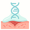
A new study published in Advanced Materials (“Diamond‐Inspired DNA Hydrogel Based on Tetrahedral Framework Nucleic Acids for Burn Wound Healing”) describes a hydrogel designed to overcome those limitations. The material is made entirely from DNA nanostructures called tetrahedral framework nucleic acids, or TFNAs. These are synthetic, self-assembling pyramids composed of single strands of DNA. Each TFNA has four outward-facing overhangs known as sticky ends. When two complementary types, named Ta and Ta*, are mixed together, they connect at these sticky ends to form a continuous, porous gel.
The result is a hydrogel with a highly ordered internal structure. The repeating tetrahedral arrangement resembles the atomic geometry of a diamond, which gives the material both its name and its strength. The researchers confirmed the gel’s architecture using fluorescence microscopy and scanning electron microscopy. The hydrogel forms quickly, stays stable at room temperature, and creates a dense yet permeable network.
Scheme of DNA hydrogel based on tetrahedral framework nucleic acids developed for skin regeneration. a) TDNs with two types of sticky ends were cross-linked based on complementary base pairing and formed into a DNA hydrogel named TDH. Leveraging the unique topological structure of tetrahedra, TDH exhibited enhanced mechanical strength and superior resistance to shear force and compression. b) The multifunctional TDH was characterized by efficient ROS scavenging and inhibited the ROS-inflammatory cascade via the NF-𝜅B pathway, thereby facilitating the transformation of pro-inflammatory macrophages into an anti-inflammatory phenotype. c) TDH promoted burn wound healing inmice. Single-cell sequencing revealed that TDH disrupted the ROS-inflammatory cascade, leading to the transformation of pro-inflammatory macrophages into an anti-inflammatory phenotype in vivo. This phenotypic change influenced cellular communication between macrophages, fibroblasts, and keratinocytes, promoting fibroblast proliferation and collagen infiltration, as well as keratinocyte proliferation and re-epithelialization. (Reprinted with permission by Wiley-VCH Verlag) (click on image to enlarge)
Mechanical testing showed that the DNA hydrogel, referred to as TDH, was much stiffer and more elastic than earlier DNA-based gels. Its storage modulus, which measures stiffness, was over eight times higher than that of a Y-shaped DNA hydrogel. TDH also resisted shear forces, recovered its shape after compression, and could be pushed through a syringe without damage. These properties suggest it could be used on joints or other areas that need to flex. The gel’s ability to switch between fluid and solid forms under pressure also allows it to be injected directly into deep or irregular wounds, where it reforms in place.
TDH also shows several biological effects that support healing. One of the most important is its ability to reduce reactive oxygen species, or ROS. These are unstable molecules produced by immune cells that can damage proteins, DNA, and membranes. While ROS help fight infection, high levels cause further inflammation. In cell culture experiments, TDH reduced intracellular ROS by about 70 percent in activated macrophages. This lowered activity in the NF kappa B signaling pathway, which controls production of inflammatory molecules like tumor necrosis factor alpha and interleukin 1 beta.
By limiting this signaling, TDH pushed macrophages away from an inflammatory state and toward a repair-focused profile. Macrophages exposed to the gel made more interleukin 10 and arginase 1, both markers of anti-inflammatory and pro-regenerative behavior. This shift is key for stopping inflammation and restarting tissue growth.
The hydrogel also helped with blood clotting. In mouse models of bleeding, including tail amputation and liver injury, TDH stopped bleeding faster and more effectively than a commercial gelatin sponge. It absorbed fluid, stuck to wet tissue, and helped clots form. These traits may make it useful when bleeding control and tissue healing are both needed.
The researchers tested the hydrogel’s safety by injecting it under the skin in mice. It caused no tissue damage or inflammation. Within four days, the material broke down into short DNA strands that were cleared from the body. These fragments didn’t trigger immune responses or accumulate in organs.
To test its healing ability, the team applied TDH to full-thickness burn wounds on mice. They compared these wounds to others treated with a standard burn ointment or with single-stranded DNA. TDH clearly sped up healing. After 14 days, wounds treated with TDH had nearly closed. Hair follicles and blood vessels had returned, and collagen fibers were better aligned. Tissue samples showed lower levels of inflammatory cytokines and higher levels of vascular endothelial growth factor, which supports blood vessel formation.
The researchers also looked at how TDH affected healing at the cellular level. They used single cell RNA sequencing to measure gene expression in thousands of cells from the wound area. TDH changed the types of cells present during healing. It reduced the number of neutrophils and inflammatory macrophages and increased fibroblasts, keratinocytes, and endothelial cells. These are the cells responsible for building new tissue and restoring skin.
Macrophages were sorted into four subtypes based on their gene activity. TDH lowered the number of inflammatory types and raised the number linked to repair. Using a method called RNA velocity, the researchers found that TDH guided macrophages to develop into pro-regenerative types rather than inflammatory ones.
TDH also affected fibroblasts, the cells that build connective tissue. It increased the number of fibroblasts making alpha smooth muscle actin and types I and III collagen. These are signs that fibroblasts are actively rebuilding tissue. The gel also turned on signaling through transforming growth factor beta and insulin like growth factor 1, which both help fibroblasts function during wound healing.
In keratinocytes, the cells that form the outer layer of skin, TDH encouraged a shift toward states that help with movement and division. These cells became more likely to undergo epithelial to mesenchymal transition, which lets them move into the wound and help close it. TDH also reduced interleukin 1 in these cells, which helped lower inflammation. Signals between keratinocytes and immune cells increased through growth factor pathways that supported tissue coordination.
Together, the findings show that TDH works on multiple levels. It reduces oxidative stress, reshapes immune behavior, and supports the cells that rebuild skin. Unlike passive wound coverings, the hydrogel interacts with the wound environment and directs the healing process.
This study offers a different approach to wound care. Instead of simply sealing the wound, the material actively influences how cells behave. The DNA-based structure is strong, biodegradable, and biologically responsive. Its ability to support healing while managing the immune response could make it useful for treating burns that don’t respond to standard care. Further research will be needed to test this strategy in larger animals or clinical trials, but the study shows how DNA nanotechnology can do more than carry drugs. It can help heal wounds from within.
If this article was useful, support our independent nanotechnology reporting with any amount.
Your contribution funds the next explainer and keeps Nanowerk open for everyone.
For authors and communications departmentsclick to open
Lay summary
Prefilled posts
Nanowerk Newsletter
Get our Nanotechnology Spotlight updates to your inbox!
Thank you!
You have successfully joined our subscriber list.
Become a Spotlight guest author! Join our large and growing group of guest contributors. Have you just published a scientific paper or have other exciting developments to share with the nanotechnology community? Here is how to publish on nanowerk.com.