| Mar 16, 2026 |
Cryo-electron tomography shows the mitochondrial protein mHsp60 restructures itself under stress to boost folding activity, offering clues to Parkinson’s disease.
|
|
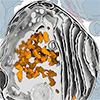
(Nanowerk News) Researchers have used cryo-electron tomography to capture the structural behavior of a key mitochondrial protein under stress conditions with near-atomic resolution. An international team led by scientists at the University Medical Center Göttingen (UMG) showed that the mitochondrial heat shock protein 60 (mHsp60), which assists other proteins in folding into their functional shapes, restructures itself during stress to boost its activity and maintain mitochondrial function.
|
|
The study, published in Science Advances (“Structural remodeling of the mitochondrial protein biogenesis machinery under proteostatic stress”), could advance understanding of neurodegenerative diseases such as Parkinson’s disease.
|
Key Findings
- The protein mHsp60 remodels its three-dimensional structure under stress, increasing its folding activity to sustain mitochondrial health.
- Cryo-electron tomography captured these structural changes directly inside human cells at near-atomic precision.
- Stressed mitochondria reduce protein translation while ramping up protein folding as part of a coordinated quality-control response.
|
|
Mitochondria generate most of a cell’s energy supply. When these organelles malfunction, tissues that consume large amounts of energy are affected first. Muscles and the brain fall into this category, and within the brain, dopaminergic neurons are especially vulnerable. Located primarily in the midbrain, these cells produce dopamine, a neurotransmitter involved in movement, motivation, mood, and drive.
|
|
In Parkinson’s disease, dopaminergic neurons progressively die, though the exact reasons remain unclear. More than 10 million patients worldwide are affected, according to the umbrella organization Parkinson’s Europe. One explanation is that the extreme energy needs of these neurons place their mitochondria under constant pressure, eventually causing them to fail. Conversely, abnormally robust mitochondrial performance may help cancer cells proliferate. Both scenarios underscore why the molecular mechanisms governing mitochondrial fitness matter across multiple disease contexts.
|
|
“We carried out a stress test within the cell’s energy factories, to analyze the molecular mechanisms of quality control and their weaknesses,” says Prof. Dr. Rubén Fernández-Busnadiego, last author of the study and head of the Structural Cell Biology group at UMG’s Department of Neuropathology. “We are particularly interested in deciphering the link between cellular stress, protein misfolding and severe neurodegenerative diseases.”
|
|
Until now, available technology could not resolve these processes at sufficient detail inside cells. Fernández-Busnadiego’s team, working within the Göttingen Cluster of Excellence “Multiscale Bioimaging” (MBExC), addressed this gap with cryo-electron tomography. The technique flash-freezes cells in an ultrafast process, preserving them in a near-native state and enabling three-dimensional imaging at near-atomic resolution.
|
|
The researchers exposed human cells to a chemical substance that causes misfolded, inactive proteins to accumulate inside mitochondria. In response, the cells produced additional mHsp60 complexes. These molecular machines encapsulate unfolded proteins within a protective barrel-shaped structure, guiding them toward their correct three-dimensional form. The cryo-electron tomography data showed how these barrels assemble inside human cells, and revealed that stress shifts the architecture of mHsp60 in a way that allows it to fold proteins more efficiently and at higher capacity.
|
|
Beyond the stress response, the imaging data also provided a detailed view of the normal functional cycle of mHsp60. The team carried out this structural and functional analysis in close collaboration with researchers from the Biofisika Institute in Spain, Tel Aviv University in Israel, and the University of Dundee in the United Kingdom. Next, the group plans to examine how this cycle breaks down under pathological conditions, including in the context of Parkinson’s disease.
|
|
Kenneth Ehses, postdoctoral researcher at UMG’s Department of Neuropathology, first author of the study, and member of the MBExC’s Hertha-Sponer-College teaching and training platform, said: “Cryo-electron tomography technology allows us to study protein complexes directly within their native cellular environment, enabling us to investigate possible mechanisms for the development of diseases. The findings could contribute to the development of new treatment strategies for neurodegenerative diseases such as Parkinson´s Disease.”
|
|
The Göttingen Cluster of Excellence MBExC, established in January 2019 under the Excellence Strategy of the German federal and state governments, studies electrically active heart and nerve cells from the molecular to the organ level. Its imaging toolkit includes optical nanoscopy, X-ray imaging, and electron tomography. Multiple university and non-university partners across the Göttingen Campus contribute to the cluster’s central aim of connecting basic and clinical research on heart and brain diseases while developing new analytical methods.
|