Crystalline polymers assembled inside tumor cell lysosomes rupture membranes, release iron, and trigger immune-activating ferroptosis that shrinks tumors in mice.
(Nanowerk Spotlight) Cells produce natural polymers such as DNA, proteins, and sugars to sustain life. Synthetic polymers can offer properties they lack: customizable architecture, tunable porosity, built-in fluorescence. Assembling them directly inside a living cell rather than in a flask could enable precise manipulation of cellular behavior that pre-fabricated materials cannot achieve. But the cell interior is aqueous, crowded, and chemically hostile to most polymerization reactions.
Several groups have nonetheless triggered simple polymer formation within cells. The products, however, have always been amorphous, lacking internal atomic order. Without that order, they are difficult to characterize and nearly impossible to engineer for predictable biological effects.
Crystalline polymers would solve both problems. Yet coaxing molecules to crystallize inside a cell has remained out of reach. The reactions that produce crystalline frameworks typically demand high temperatures, toxic organic solvents, vacuum conditions, and reaction times of days. No living cell survives those conditions.
The polymers are covalent organic frameworks (COFs), porous structures in which organic building blocks link through strong covalent bonds into repeating lattices. First described in 2005, COFs have attracted interest in catalysis, gas storage, and biomedicine, but their synthesis has historically required conditions incompatible with biology.
The research team, based primarily at the University of Macau, targeted lysosomes, the acidic digestive compartments that break down cellular waste. Lysosomes maintain an internal pH of roughly 4.5 to 5.0. That natural acidity can catalyze imine condensation, a reaction joining an amine group to an aldehyde group to form a carbon-nitrogen double bond.
The team selected two small organic monomers: TAPB, carrying three amine groups around a central benzene ring, and DMTP, a dialdehyde. Mixed in mildly acidic phosphate buffer at 37 °C with the nonionic surfactant Tween-80 as a solubilizer, these monomers polymerized and crystallized into a COF designated UMCOF1. Powder X-ray diffraction confirmed the atomic order, and computational modeling matched the data to a layered honeycomb structure.
Synthesis of UMCOF1 in a phosphate buffer. (A) Aqueous-phase synthesis and crystal structure of the bisimine model compound (CDCC 2361348). (B) Synthesis and structure of UMCOF1. (C) UV–vis spectrum of UMCOF1. Inset: digital photographs of the reaction bottle charged with the phosphate buffer (10 mM, pH 5.0) containing TAPB (50 µM), DMTP (75 µM), and Tween-80 (0.0213 vol. %) at 37°C for 24 h. (D) Time-dependent absorbance of the reaction buffer solutions (pH = 4.0–8.0) at 427 nm. Data are expressed as mean ± SD; n = 4 independent experiments. (E) PXRD patterns of the products isolated at different reaction times. (F) Experimental (grey dots), Pawley-refined (red), and simulated (blue) PXRD patterns; difference plot (green); and the Bragg positions (brown) of the UMCOF1 powder. Inset: amplified curves in the range of 2θ = 2°–11°. (Image: Reproduced from DOI:10.1002/adma.202510663, CC BY) (click on image to enlarge)
The reaction reached equilibrium within 6 hours at pH 4.0 to 5.0 but stalled at neutral pH. After 24 hours, the isolated product appeared as an insoluble yellow powder in 76 % yield. Four additional COF structures prepared under similar aqueous conditions confirmed the method’s generality.
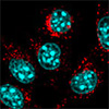
Translating this chemistry into cells, the team incubated 4T1 mouse breast cancer cells with both monomers. The molecules entered the cells and accumulated in lysosomes, where acidity drove polymerization. UMCOF1 emits green fluorescence when excited by a 405 nm laser, enabling real-time tracking. The fluorescent signal overlapped strongly with a lysosome-specific dye.
Pretreatment with Bafilomycin A1, a drug that neutralizes lysosomal acidity, cut fluorescence to roughly 28 % of untreated levels, confirming the acidic environment was essential. Transmission electron microscopy revealed spherical nanoparticles approximately 120 nm across within lysosomes. High-resolution imaging showed an interplanar spacing of 3.5 Å consistent with the expected π–π stacking of UMCOF1 layers. X-ray diffraction of material extracted from whole-cell lysates verified the material’s atomic order.
Building a crystalline polymer inside a lysosome proved destructive. COF formation ruptured lysosomal membranes, spilling contents including stored ferrous iron into the cytoplasm. The surge in free iron catalyzed lipid peroxidation, a chain reaction in which reactive oxygen species attack the fatty acid tails of membrane lipids. This cascade triggered ferroptosis, a form of regulated cell death defined by iron dependence and runaway lipid damage, distinct from apoptosis, the orderly self-destruction program cells typically use.
The concentration required to kill half the cancer cells was approximately 44.3 µM (TAPB equivalent). Established ferroptosis inhibitors reversed the effect, as did the iron chelator deferoxamine. Individual monomers alone and pre-synthesized UMCOF1 nanoparticles added from outside caused no toxicity. Assembling the framework inside lysosomes, not mere chemical exposure, drove cell death.
Whether ferroptosis alerts the immune system or suppresses it remains actively debated. The team compared their approach with RSL3, a ferroptosis inducer that blocks GPX4, an enzyme that normally neutralizes lipid peroxides to protect membranes. RSL3 triggers lipid damage primarily in the endoplasmic reticulum, the cell’s protein-processing network.
Both treatments released molecular distress signals known as damage-associated molecular patterns, including ATP and the protein HMGB1. But COF-induced ferroptosis released these signals more slowly and continuously. It also drove the protein calreticulin to the outer cell surface, a recognized flag that marks dying cells for immune engulfment.
RSL3-treated cells upregulated calreticulin only internally. When culture supernatants from COF-treated cancer cells contacted macrophages, those immune cells became activated, releasing inflammatory cytokines. Supernatants from RSL3-treated cells did not produce this effect.
In mouse tumor models, intratumoral injection of the two monomers into 4T1 breast tumors shrank tumor weight to 19 % of untreated animals after 15 days, with no significant weight loss or organ toxicity. Classically activated splenic macrophages increased roughly 6.6-fold. Mature dendritic cells rose nearly 5-fold. The ratio of cytotoxic to helper T cells climbed significantly.
When combined with the immune-stimulating agent R848 in a bilateral tumor model, the treatment inhibited not only the injected primary tumor but also distant, untreated tumors, a systemic response known as the abscopal effect. A tumor vaccine prepared from lysates of COF-treated cells delayed tumor formation in immunocompetent mice, with half showing no tumor growth, but failed entirely in T cell-deficient nude mice, underscoring adaptive immunity’s central role.
The study carries acknowledged limitations. Intracellular COF synthesis also killed normal cell lines in culture, showing no inherent tumor selectivity. Intratumoral injection limits healthy tissue exposure, but systemic delivery would require tumor-targeting strategies. The long-term fate of monomers escaping the tumor site also remains unstudied.
By converting lysosomes into nanoscale reaction vessels, this work demonstrated that a crystalline, porous polymer can self-assemble from simple precursors under conditions a living cell naturally provides. The framework’s built-in fluorescence and defined structure allowed direct verification that truly ordered material had formed, addressing a persistent challenge in intracellular polymerization research.
Whether the approach can develop into a viable cancer therapy remains uncertain, but it establishes a new category of interaction between synthetic chemistry and cellular biology.
For authors and communications departmentsclick to open
Lay summary
Prefilled posts
Nanowerk Newsletter
Get our Nanotechnology Spotlight updates to your inbox!
Thank you!
You have successfully joined our subscriber list.
Become a Spotlight guest author! Join our large and growing group of guest contributors. Have you just published a scientific paper or have other exciting developments to share with the nanotechnology community? Here is how to publish on nanowerk.com.