| Jan 19, 2026 |
By combining atomic force microscopy (AFM) with a Hadamard product-based image reconstruction algorithm, scientists successfully visualized the nanoscopic dynamics of membrane rafts in live cells.
(Nanowerk News) Membrane rafts are nanometer-scale structures rich in cholesterol and sphingolipids, believed to serve as vital platforms for cell signaling, viral entry, and cancer metastasis. Since the concept emerged in the 1990s, the existence and behavior of these lipid domains have been intensely debated.
|
|
Conventional fluorescence microscopy, typically performed on fixed and stained cells, could not capture features that are only tens of nanometers wide and change within seconds. As a result, even the question of whether such rafts exist on live membranes remained unresolved for decades.
|
|
Led by PhD candidates Ms. Hsiang-Ling Chuang and Ms. Yu-Chen Fa at National Taiwan University, the research team employed high-resolution AFM in conjunction with Hadamard product–based image processing to record, in real time, the formation, fusion, and dissolution of membrane rafts on live cell surfaces. Using C-Laurdan phase-sensitive dyes and integrin co-localization imaging, they confirmed that the nanoscale domains observed by AFM indeed correspond to membrane raft structures.
|
|
This discovery, published in Science Advances (“Visualizing dynamics of membrane rafts on live cells”), marks the first direct visualization of lipid raft dynamics in live cells.
|
 |
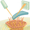
| Illustration of membrane rafts organized by theoretical mechanisms. (Image: National Taiwan University)
|
|
The study revealed that these rafts are highly dynamic liquid-ordered (Lo) regions, typically 10 to 200 nanometers in diameter, continuously reorganizing through interactions among lipids, proteins, and the cytoskeleton.
|
|
According to chemist Prof. Chun-hsien Chen, AFM provides nanoscale measurements of surface height and stiffness on live cells, while Hadamard product analysis effectively suppresses irrelevant background signals, enhancing the visibility of raft-related features. “This combination allows us to identify subtle, transient signals that conventional optical techniques could never resolve,” Prof. Chen explained.
|
|
Building on the current understanding of membrane rafts, co-corresponding author Prof. Ja-an Annie Ho noted, “This technology, which enables the visualization of membrane dynamics in real time, could become a rapid screening platform for drug discovery.”
|
|
Integrating chemistry, biophysics, and biochemical technology, this interdisciplinary study opens a new window into the nanoscale organization of live membranes, offering powerful new tools for drug development and disease mechanism research.
|