An implantable scaffold and magnetic probes capture circulating tumor cells directly in the bloodstream, eliminating over 90% of captured cells in rabbits and goats.
(Nanowerk Spotlight) A single cancer cell breaks free from a tumor, squeezes through the wall of a nearby blood vessel, and enters the rushing current of the bloodstream. Within seconds, it is hurtling through the body at speeds that would carry it from head to toe in under a minute. Most of these escaped cells will die, battered by shear forces or destroyed by immune sentinels. But a few will survive, lodging in distant organs and seeding the secondary tumors that cause more than 90% of cancer deaths. These travelers are called circulating tumor cells (CTCs), and they represent both a tantalizing target and a formidable adversary.
The logic is straightforward: intercept these cells during their journey and destroy them before they can establish new colonies. In the laboratory, this idea has shown real promise. Microfluidic chips and sophisticated sorting systems can capture CTCs with efficiencies exceeding 90%. But the living bloodstream is a far more hostile environment. Blood flows fast, immune cells compete for space, and CTCs are extraordinarily rare, sometimes just a handful among billions of normal cells.
Early attempts to capture CTCs inside the body involved threading antibody-coated wires into veins. The approach proved that in vivo capture was possible, but limited surface area and poor contact with fast-moving blood produced meager results. External loop systems that divert blood through a capture module before returning it to the body have also been tested, but processing large volumes at realistic flow rates remains impractical. I
mplantable magnetic devices and therapeutic catheters have shown more promise in animal models, yet none has achieved both high capture efficiency and selective elimination of captured cells without collateral damage to healthy tissue. Before this work, the best reported in vivo capture efficiency stood at 64.2%, and most approaches fell far short.
A study published in Advanced Materials (“Thread‐Designed Vascular Scaffold with Magneto‐Optical Probes Capture and Elimination of Circulating Tumor Cells In Vivo”) by researchers in China, takes a different approach. The team developed an integrated system combining a specially designed implantable vascular scaffold with magneto-optical probes that capture and destroy CTCs directly within blood vessels. In experiments on rabbits and goats, the platform achieved capture efficiencies of 60.3% and 54.7% respectively, with post-capture elimination rates exceeding 90%.
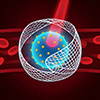
Schematic illustration of in vivo circulating tumor cells (CTCs) capture and in situ elimination using the HMs-coated magneto-optical probe and a threaded vascular scaffold: (A) Synthesis process of the magneto-optical probe; (B) Fabrication and in vivo implantation of the threaded vascular scaffold; (C) Under magnetic guidance, CTCs specifically targeted by the probe are directed to accumulate on the scaffold’s inner surface, while normal blood cells pass through the central lumen, enabling efficient CTCs capture and enrichment at the scaffold site; (D) Under NIR light irradiation, specific elimination of CTCs is achieved under high blood flow velocity in vivo. (Image: Reproduced with permission from Wiley-VCH Verlag) (click on image to enlarge)
The system has two main components. The first is a threaded vascular scaffold made from polylactic acid nanofibers, fabricated using a custom electrospinning technique. The scaffold’s inner surface features a helical, thread-like structure engineered to manipulate blood flow dynamics.
Finite element simulations showed that this threaded design alters radial velocity distribution inside the scaffold, slowing fluid near the walls while maintaining faster flow along the central axis. This creates a cell-enrichment zone: CTCs are funneled toward the scaffold’s inner surface where they can be captured, while normal blood cells pass through the center relatively unimpeded. The design also subjects captured cells to lower shear forces, helping retain them once bound.
The second component consists of hybrid membrane-modified magnetic beads, designated HM-MBs. These porous iron oxide nanoparticles measure approximately 173 nm in diameter and carry a payload of indocyanine green, a near-infrared-absorbing dye that the FDA has approved for medical imaging and other clinical uses.
A fusion of tumor cell membranes and white blood cell membranes coats each particle. The tumor membrane enables the beads to recognize and bind specifically to CTCs through homotypic targeting, a process in which surface proteins on the coating interact with matching proteins on circulating cancer cells. The white blood cell membrane helps the beads evade immune detection and reduces nonspecific binding to normal blood components.
Once injected, the HM-MBs circulate through the bloodstream and latch onto CTCs. An external magnetic field then draws the labeled cells toward the implanted scaffold, concentrating them at the capture site. Near-infrared laser light triggers both photodynamic and photothermal effects from the indocyanine green, generating reactive oxygen species and localized heat that destroy the captured cancer cells. Because the therapeutic agents attach directly to CTCs rather than disperse throughout the scaffold, the destruction is selective and spares unlabeled healthy cells.
The researchers built an additional safety mechanism into the nanoparticles. During synthesis, they incorporated L-arginine into the porous iron oxide structure. When near-infrared light generates reactive oxygen species, these molecules convert the L-arginine into nitric oxide. This gas is a natural signaling molecule that dilates blood vessels and suppresses inflammation. The controlled release helps counteract the vasoconstriction and inflammatory responses that intravascular procedures can provoke, reducing the risk of complications during treatment.
In vitro tests using a closed-loop circulation system demonstrated capture efficiencies up to 83.7% for scaffolds with a 2 mm diameter, with capture purity reaching 98%. In rabbits, scaffolds implanted in the abdominal wall vein captured 60.3% of injected tumor cells under magnetic guidance, and subsequent laser treatment eliminated 92% of those captured.
The team also tested the system in goats. These animals offer practical advantages: their gentle temperament facilitates postoperative observation, and their jugular veins, measuring approximately 5.4 mm in outer diameter, approximate human peripheral vessels more closely than the ear veins of rabbits or pigs used in earlier studies. In goats, capture efficiency reached 54.7%, with an elimination rate of 90.5%.
Safety assessments proved encouraging. Blood tests on rabbits at intervals after the procedure showed initial shifts in white blood cell, red blood cell, and platelet counts consistent with normal surgical inflammation. These values returned to baseline within 14 days. Liver and kidney function, coagulation markers, and immunoglobulin levels showed no significant abnormalities.
The researchers acknowledged limitations. CTC heterogeneity, the tendency of these cells to change their surface protein profiles as they circulate, may reduce targeting efficiency in some clinical scenarios. Near-infrared light penetration is limited, potentially restricting the approach to vessels accessible from the body surface. Comprehensive long-term safety studies would be necessary before clinical application.
The work represents a meaningful advance in efforts to intercept metastasis at its source. By combining biomimetic targeting, fluid dynamics engineering, and light-triggered therapy with built-in vascular protection, the system addresses obstacles that have hindered previous in vivo capture strategies. Successful demonstration in large animals with clinically relevant vessel sizes moves this technology closer to potential human application.
For authors and communications departmentsclick to open
Lay summary
Prefilled posts
Nanowerk Newsletter
Get our Nanotechnology Spotlight updates to your inbox!
Thank you!
You have successfully joined our subscriber list.
Become a Spotlight guest author! Join our large and growing group of guest contributors. Have you just published a scientific paper or have other exciting developments to share with the nanotechnology community? Here is how to publish on nanowerk.com.