Molecular coatings can slow or accelerate photon emission from near-infrared quantum dots, adding a new control handle for silicon photonics.
(Nanowerk Spotlight) Quantum dots earned much of their appeal from a simple form of control: change the size of the nanocrystal, and the color of the emitted light changes with it. That size-color relationship makes these particles useful as tunable emitters. But a light source is not defined by wavelength alone. For optical communication, sensing, and photonic circuits, the timing of photon emission can matter just as much as the color.
That second form of control is harder to engineer. The rate at which a quantum dot emits photons depends on how electrons and holes recombine inside a crystal only a few nanometers across. It can also depend on features outside the crystal, including the molecules attached to its surface and the material surrounding it. These external influences are easy to treat as secondary details, but they can reshape the emitter’s behavior.
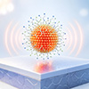
Figure 1: Quantum dot (QD) nanocrystals with varying sizes and surface coatings. Especially smaller nanocrystals exhibit a strong influence of the so-called Stark effect that is caused by electric fields. (Image: Courtesy of the researchers)
The work shows that molecular coatings on the dot surface can strongly alter how fast the particles emit photons. The researchers propose that some ligands generate local electric fields that separate electron and hole wave functions inside smaller dots, slowing radiative recombination.
The same study also shows that placing the dots on silicon can accelerate emission, giving the surrounding optical environment its own role in rate control. The result reframes near-infrared quantum dots for silicon photonics. Choosing the right particle size remains essential because it sets much of the emission wavelength. But size alone does not determine whether the emitter will behave well in a device.
The group studied commercially available lead sulfide quantum dots because they emit in the near-infrared, including wavelengths compatible with silicon photonic structures and optical communication bands. Silicon can guide light efficiently at these wavelengths, but it is a poor light emitter. Adding nanoscale emitters to silicon structures offers one route toward active photonic devices, provided the emitters can be tuned in both wavelength and emission dynamics.
The quantum dots carried one of two surface chemistries. Some were capped with oleic acid, a common ligand that stabilizes particles in nonpolar solvents such as toluene. Others were capped with polyethylene glycol ligands ending in amine groups, which support use in more polar environments, including water. This comparison allowed the researchers to separate the role of the inorganic lead sulfide core from the role of the molecular shell around it.
“Our team measured many different sizes of quantum dots suspended in various liquids,” says Andreas Schulz, first author of the study. “We found that the special coating on the quantum dots creates tiny electric fields. These fields change the results of our measurements, showing how important the surface chemistry is when studying semiconductor nanocrystals.”
The expected size effect appeared first. Emission spectra showed broad near-infrared peaks whose positions shifted with nanocrystal diameter. Smaller dots emitted at higher photon energies, while larger dots emitted at lower photon energies. This behavior follows from quantum confinement, where electrons and holes occupy size-dependent energy states because the crystal confines them within only a few nanometers.
The more revealing measurements examined what happened after a short pulse of excitation. If every dot in a sample emitted photons at the same rate, the light intensity would decay with a single characteristic lifetime. Some oleic-acid-coated dots came close to that behavior near their emission peaks. Other samples did not. Their decay curves showed that the ensemble contained a spread of emission rates rather than one uniform value.
That spread is not a technical detail. A single average lifetime can blur the distinction between a nearly uniform emitter and an ensemble whose surface chemistry creates a range of recombination pathways. To capture that variation, the researchers modeled the measurements with a log-normal distribution of decay rates. This approach described both narrow and broad distributions without forcing the dots into an artificial single-lifetime picture.
The strongest contrast emerged when the surface coatings were compared. Oleic-acid-coated dots in toluene showed only modest changes in their most frequent decay rate across the measured range. Their behavior matched a two-level exciton model better than an atom-like emitter model. In other words, the dots behaved less like artificial atoms and more like tiny semiconductors in which an electron-hole pair recombines across the confined crystal.
Polyethylene-glycol-coated dots departed from that pattern. For dots smaller than about 4.4 nm, the most frequent decay rate decreased sharply. The paper proposes that the ligand shell causes this slowdown by generating local electric fields. In water, amine end groups can become protonated and carry positive charge. In chloroform, the same groups are neutral but polar. Both situations can produce an electric influence near the semiconductor core.
The physical picture is direct. A smaller dot has more surface area relative to its volume, so the chemistry at the surface can affect a larger fraction of the nanocrystal. An electric field from the ligand shell can enhance the quantum-confined Stark effect, which shifts electron and hole wave functions away from one another. With less overlap between those wave functions, radiative recombination becomes less likely, and photon emission slows.
“We observed a pronounced quantum-confined Stark effect in some of our quantum dots, arising from the electric fields generated at the nanocrystal surface,” says Christian Blum. He notes that ligand-induced Stark shifts could be useful for developing sensitive nanoscale sensors, including possible applications in biophysical imaging and single-molecule electrophysiology.
The paper treats this explanation carefully. It also weighs other possibilities, including solvent polarity, quenching, solvent resonances, acoustic effects, and trapping. Several alternatives do not fit the direction of the measured change. Common quenching pathways, for example, would tend to increase nonradiative decay and therefore raise the total decay rate rather than reduce it.
This ligand effect changes how the surface coating should be understood. In many quantum dot applications, ligands are chosen mainly to keep particles dispersed, prevent aggregation, or attach the nanocrystals to other materials. Here, the surrounding molecules appear to influence the electronic overlap that controls emission from the core. The coating becomes part of the optical design, not merely a chemical support layer.
Figure 2. Transmission electron micrograph (TEM) of PbS QDots, covered with −PEG−NH2 ligands. Yellow circles mark the position of individual QDots. The scale bar represents 10 nm. Inset: zoom-in of two PbS quantum dots revealing crystal planes. The scale bar represents 5 nm. (Image: Courtesy of the researchers)
The silicon result adds a second control handle. When quantum dots were deposited on a silicon surface, their emission rates increased by roughly 5-fold to 10-fold compared with dots in suspension. The paper attributes this increase to the higher dielectric function near silicon. Silicon changes the electromagnetic environment around the emitter, which can increase the measured emission rate.
“Our results reveal exciting new ways to dynamically control light at the nanoscale,” says Willem Vos. He points to possible applications in ultrafast optical modulators, sensitive electric-field sensors, and bio-photonic devices. In device terms, the quantum dot should not be treated as an isolated light source whose behavior is fixed once its size and composition are chosen. Ligands can influence internal recombination through local fields, while silicon can alter the external optical environment.
The work does not present a completed quantum-dot light source for integrated silicon photonics. Its value lies in identifying which variables control different parts of the emitter’s behavior. Size remains the main handle for wavelength. Surface chemistry and dielectric environment provide handles for timing. The lesson is that color tuning is only the starting point. For near-infrared quantum dots in silicon photonics, the molecules and materials around the nanocrystal may be just as important for controlling when photons are released.
For authors and communications departmentsclick to open
Lay summary
Prefilled posts
Nanowerk Newsletter
Get our Nanotechnology Spotlight updates to your inbox!
Thank you!
You have successfully joined our subscriber list.
Become a Spotlight guest author! Join our large and growing group of guest contributors. Have you just published a scientific paper or have other exciting developments to share with the nanotechnology community? Here is how to publish on nanowerk.com.