A biodegradable ionic hydrogel battery modeled on electric eel organs powers a nerve conduit that accelerates peripheral nerve regeneration without external energy.
(Nanowerk Spotlight) Every nerve signal in the body is, at its core, an electrical event. When a peripheral nerve is severed, the biological wiring that carries those signals goes dark. Restoring it requires more than a physical bridge between the cut ends. The regenerating tissue also needs electrical cues to guide new axons, activate the support cells that wrap them in insulating myelin, and recruit blood vessels to feed the growing fibers.
Applying electricity to injured nerves speeds regrowth. Clinical and laboratory studies have confirmed this repeatedly. The problem lies not in the principle but in the delivery. External stimulators require wires or batteries that risk infection. Piezoelectric materials are rigid and can damage soft tissue.
Organic piezoelectric films are typically non-degradable, forcing a second surgery for removal. Magnetically driven systems add bulk and cost. Each approach solves one problem while creating another.
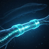
Electric eels offer a biological template for a different kind of power source. Their electric organs contain thousands of disc-shaped cells called electrocytes, stacked in series. Each electrocyte generates roughly 150 mV by shuttling sodium and potassium ions across selective membranes. Stacked together, these cells produce hundreds of volts on demand.
Researchers have previously drawn on this architecture to build soft, stretchable gel-based batteries, but those prototypes used non-degradable polymers, limiting their usefulness inside the body.
The device consists of three concentric layers. An inner tube of polycaprolactone blended with methacrylated chitosan forms the structural conduit. A middle layer houses the ionic hydrogel battery. An outer electrospun membrane seals the assembly and isolates it from surrounding tissue.
(A) Anatomical structure of an electric eel. (B) Mechanism of voltage generation in electrocytes. (Image: Adapted with permission from Wiley-VCH Verlag) (click on image to enlarge)
The battery’s repeating unit mirrors the electrocyte stack. Four hydrogel types alternate in sequence: a high-salinity gel, a cation-selective gel, a low-salinity gel, and an anion-selective gel. When these layers contact one another, ions migrate along the concentration gradient through the selective membranes. This process, reverse electrodialysis, generates a voltage of up to 150 mV per unit.
The researchers chose chitosan and hydroxyethyl cellulose as base polymers because both are naturally derived, biodegradable, and biocompatible. Chemical modification with methacrylate groups allowed UV-triggered crosslinking, producing stable gel networks. Connecting multiple battery units in series or parallel scaled the output proportionally.
Magnesium chloride served as the electrolyte. The team selected it over sodium chloride for its higher ionic strength and over other divalent salts for its low cytotoxicity. Prior work has shown that magnesium ions activate the PI3K/Akt signaling pathway, which promotes axonal growth, giving the electrolyte a dual role as both charge carrier and bioactive agent.
The next challenge was confirming that the battery could sustain useful output long enough to support nerve regeneration. Over 30 days, voltage dropped from 275 mV to 61 mV as the ionic gradient gradually equilibrated. Even at 61 mV, the output fell within the range needed to recruit cells and growth factors during the early repair window. The hydrogel scaffold degraded by 30 to 42 percent over three months, maintaining structural support throughout the most vulnerable period of tissue regrowth.
Testing the device in a rat sciatic nerve injury model provided the strongest evidence of its therapeutic value. The team compared four groups: an empty conduit control, a hydrogel-only conduit without electrical output, the electrically active conduit, and an autograft, the clinical gold standard. After four weeks, the electrically stimulated group showed markedly higher levels of the axonal marker NF200 and the Schwann cell marker S100, second only to the autograft group.
By 12 weeks, dense and well-myelinated nerve tissue filled the stimulated conduits. Schwann cell density and myelin coverage exceeded those of the control and hydrogel-only groups. Electron microscopy revealed thicker myelin sheaths and larger axon diameters, hallmarks of more mature regenerated fibers. The gastrocnemius muscle on the injured side retained more mass, indicating reduced atrophy from faster nerve reinnervation.
Functional measures matched the structural improvements. Nerve conduction velocity in the stimulated group reached 52.8 m/s, representing a 79.6 percent recovery rate compared with 46.4 percent in the untreated group. The sciatic functional index, a measure of walking ability, approached autograft-level performance.
The biological mechanisms behind these gains came into focus through transcriptomic analysis of nerve tissue at one week. Electrical stimulation upregulated genes linked to M2 macrophage polarization, a phenotype associated with tissue repair rather than inflammation. At the same time, the treatment suppressed inflammatory and fibrotic pathways, including TNF, NF-κB, and IL-17 signaling.
The stimulated tissue also showed elevated VEGF and CD31 expression, confirming that the weak current promoted blood vessel formation. New vasculature supplies oxygen to regenerating axons and provides physical tracks for Schwann cell migration. These vascular and immunomodulatory effects distinguish this device from passive scaffold-based approaches to nerve repair, which lack an intrinsic mechanism for electrical stimulation.
Organ-level safety data at 12 weeks revealed no detectable damage to the heart, liver, spleen, lungs, or kidneys. Magnesium ion concentrations in tissues remained within normal physiological ranges, and chitosan degradation products appeared only at trace levels or not at all.
The authors acknowledge several limitations. Validation in large animal models, particularly primates, must precede clinical translation. Battery configuration will likely require customization for different injury types and gap lengths. The inherent voltage decay concentrates the strongest stimulation in the early repair phase, which may or may not suit all clinical scenarios. Developing external circuit control to fine-tune discharge parameters represents a logical next step. Even so, the combination of full biodegradability, self-powered electrical output, anti-inflammatory modulation, and pro-angiogenic activity in a single implantable conduit establishes a new design framework for peripheral nerve regeneration therapy.
For authors and communications departmentsclick to open
Lay summary
Prefilled posts
Nanowerk Newsletter
Get our Nanotechnology Spotlight updates to your inbox!
Thank you!
You have successfully joined our subscriber list.
Become a Spotlight guest author! Join our large and growing group of guest contributors. Have you just published a scientific paper or have other exciting developments to share with the nanotechnology community? Here is how to publish on nanowerk.com.