| Apr 02, 2026 |
A new plasmonic fiber probe uses linearly polarized light and energy recycling for broadband nanofocusing with resolution rivaling atomic force microscopy.
(Nanowerk News) A fiber-based plasmonic probe that operates with ordinary linearly polarized light has resolved features nearly matching atomic force microscopy precision. Developed by researchers at Xi’an Jiaotong University and published in Microsystems & Nanoengineering (“Broadband plasmon modulation and high-intensity nanofocusing for high-resolution nanoscale imaging using Fabry–Pérot probes”), the double-slit plasmonic platform probe, or DSPP, combines energy recycling through Fabry–Pérot interference with a precision-fabricated tip to deliver strong, stable nanofocusing without the complex illumination that existing designs require.
|
Key Findings
- At 633 nm wavelength, the DSPP tip generated roughly six times the electric field strength of a comparable asymmetric double-slit probe.
- Stable nanofocusing held across a 580 to 800 nm range, with the largest gains at shorter wavelengths where plasmonic losses are typically most damaging.
- The probe resolved a 28.6 nm slit in optical imaging, closely matching the 28.2 nm measurement obtained by atomic force microscopy, while confocal microscopy produced only a blurred outline.
|
|
Conventional plasmonic probes squeeze light below the diffraction limit by converting photons into surface plasmon polaritons, collective oscillations of electrons and electromagnetic fields that travel along metal surfaces. This confinement makes them valuable for super-resolution imaging, spectroscopy, sensing, and nanoscale detection.
|
|
In practice, however, most designs depend on radially polarized illumination, which demands specialized optics and careful alignment. Propagation losses, modest field enhancement at the apex, and difficulty fabricating consistent sub-20 nm tips further erode performance and reproducibility.
|
 |
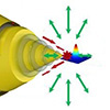
| Plasmonic Fiber Probe Enables Sharper Nanoimaging. This schematic illustrates the double-slit plasmonic platform-based fiber probe developed for broadband, high-intensity nanofocusing and high-resolution optical imaging. By combining phase interference and plasmonic field confinement, the probe enhances light concentration at the tip and enables nanoscale imaging with resolution beyond the diffraction limit. (Image: Reproduced from DOI:10.1038/s41378-026-01197-1, CC BY)
|
|
The DSPP addresses these constraints through two integrated mechanisms. A pair of slits etched into the fiber converts linearly polarized input directly into counter-propagating surface plasmon polaritons, eliminating the need for radial polarization altogether. Behind the tip, a platform-shaped reflective surface bounces a portion of the plasmon energy back toward the apex. The returning waves interfere constructively with the forward-traveling plasmons, amplifying the electric field at the tip without any increase in input power.
|
|
Tip fabrication proved equally important. The team used a focused ion beam sleeve-ring etching strategy to carve the front cone, achieving a tip radius of approximately 15 nm. This method improved tip curvature by more than an order of magnitude over conventional approaches, yielding both a sharper apex and more uniform geometry from probe to probe.
|
|
Simulations and experiments confirmed substantial performance gains. The six-fold electric field enhancement over an asymmetric double-slit reference probe translated into a far more intense optical signal at the tip. Broadband stability was equally notable: the DSPP maintained consistent nanofocusing from 580 to 800 nm, and the improvement margin widened at shorter wavelengths, precisely the spectral region where plasmonic losses normally suppress signal most aggressively.
|
|
Imaging tests demonstrated the practical impact of these gains. When scanned across a narrow slit, the DSPP returned a width measurement of 28.6 nm, virtually identical to the 28.2 nm value recorded by atomic force microscopy. Confocal microscopy applied to the same feature could not resolve it at all. Beyond raw resolution, the probe captured both surface shape and optical response from deep-subwavelength structures simultaneously, providing two complementary channels of information in a single scan.
|
|
According to the authors, no single element accounts for the probe’s performance. Rather, it emerges from the interplay of simplified excitation, enhanced tip fields, broadband operation, and reproducible fabrication within one compact, fiber-integrated device. That combination lowers the practical threshold for routine use under ambient laboratory conditions, where alignment tolerance and signal consistency often matter as much as peak resolution.
|
|
The intense, tightly confined fields and wide operating bandwidth suit the probe to applications including single-molecule detection, nanoscale spectroscopy, biological cell imaging, subwavelength lithography, and defect inspection on optical chips. The fabrication method itself may carry additional weight: by delivering tighter structural control over tip geometry, it could help shift advanced plasmonic probes from one-off laboratory prototypes toward repeatable, standardized production.
|