Custom polymer structures can now be 3D printed inside living cells using laser-based fabrication, opening paths to intracellular sensors, cell tracking tags, and embedded microlasers.
(Nanowerk Spotlight) A human cell is roughly 20 micrometers across, about one-fifth the width of a human hair. Into this space are packed thousands of proteins, organelles, and molecular machines. Placing structures inside this environment, such as sensors, mechanical probes, or tracking tags, could let researchers monitor a cell’s chemistry in real time, test how it responds to physical force, or follow individual cells as they migrate and divide.
But getting solid objects inside has proved difficult. Particles larger than roughly one micrometer cannot enter most cells. Immune cells can engulf foreign bodies through phagocytosis, but this deposits objects in membrane-bound compartments, not the cytoplasm where they could interact freely with cellular machinery.
Other techniques such as microinjection and membrane poration work for delivering molecules but have not been used to place free-standing solid structures directly into the cytosol.
Two-photon polymerization suggested another path. This technique uses a femtosecond laser to harden light-sensitive resin at a single focal point, building solid structures layer by layer with features as small as 100 nm. The method has produced micro-optics, tissue scaffolds, and structures printed inside living fruit fly embryos.
But printing inside an individual mammalian cell posed a different challenge: the laser and raw materials would need to operate within a space smaller than most printed objects themselves, without poisoning the cell or destroying its internal architecture.
A research team based in Slovenia has now demonstrated that this is possible. In a study published in the journal Advanced Materials (“Two‐Photon 3D Printing of Functional Microstructures Inside Living Cells”), the group shows that custom-shaped polymer microstructures can be fabricated inside living human cells using two-photon polymerization. The work establishes intracellular biofabrication as a new approach to engineering the cellular interior.
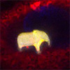
Procedure of 3D Printing in Cells. (a) Schematic representation of the printing protocol. The photoresist is injected into a cell and illuminated in a pre-designed pattern to form a polymerized structure. (b) A droplet of photoresist, injected into a live HeLa cell, dissolves almost completely in the first two hours after the injection. White arrows point to the photoresist residue approximately 2 micrometers in size, which can sometimes be observed in cells even after a long time. (c) A droplet of photoresist, injected into a live HeLa cell, is completely exposed to laser light approximately 30 min after the injection. After exposure, the photoresist polymerizes, and the droplet stops dissolving. (d) Bright-field image of a 10 micrometer elephant, printed inside a live HeLa cell. (e) Confocal image of the structure in (d). Cross-sections xz and yz clearly show that the structure is embedded in the cell, as the membrane (red) is seen covering the elephant (yellow). To improve the structure shape recognition and nucleus visibility in the xy-panel, instead of the usual confocal cross-sections the yellow and blue channels show maximum-intensity projections along the z-axis. (Image: Reproduced from DOI:10.1002/adma.202519286, CC BY) (click on image to enlarge)
The technique begins with microinjection. Using fine glass micropipettes pulled to sub-micrometer tips, the researchers delivered droplets of a commercial photoresist called IP-S into HeLa cells, a widely used human cell line. This photoresist had to meet unusual requirements: biocompatibility in both liquid and hardened states, plus slight water solubility so unpolymerized material would eventually dissolve away.
Once inside a cell, the team illuminated a photoresist droplet roughly 10 to 15 μm in diameter with a 780 nm femtosecond laser through a high-numerical-aperture microscope objective. Two-photon absorption occurred only where the laser intensity peaked, allowing precise three-dimensional patterning. The laser’s focal spot moved layer by layer through the material at 10 000 μm per second, polymerizing each designed slice in turn.
A complete 10 μm structure required 3 to 10 seconds to print. Unpolymerized photoresist then slowly dissolved over several hours, leaving only the hardened structure embedded in the cell.
Resolution inside cellular droplets approached bulk printing capabilities. The refractive index mismatch between the photoresist (n = 1.48) and surrounding cytoplasm (n = 1.36 to 1.39) causes some focal distortion, but defocusing remained below the diffraction limit of 400 nm across more than 90 percent of a typical droplet’s volume. Scanning electron microscopy confirmed walls as thin as 260 nm with sub-micrometer periodicity.
The researchers printed structures including a 10 μm elephant figure, institutional logos, hollow spheres, and woodpile lattices. Confocal microscopy confirmed these objects resided within the cell membrane, with cellular nuclei visibly deforming to accommodate the printed material.
Cell mortality matched rates seen with other established manipulation techniques. After 24 hours, roughly 55 percent of cells with printed structures were non-viable, similar to the 50 percent mortality observed after membrane puncture alone. Additionally, many cells detached from the substrate and were lost during medium exchange, a problem more pronounced in cells injected with droplets than in controls.
The researchers attribute this to membrane reshaping required to accommodate the injected material, which likely weakens cellular attachment. These results suggest membrane penetration, rather than photoresist toxicity or laser exposure, primarily contributes to cell death.
Cells containing printed structures exhibited normal morphology and continued to divide. Time-lapse imaging captured mitosis, with printed objects passing to daughter cells. However, large structures greater than 5 μm extended division time by at least one hour, indicating intracellular foreign bodies can influence cellular behavior.
The team explored several functional applications. They designed three-dimensional graphical barcodes using stacked grids of cylinders. With 61 positions available for binary encoding after accounting for structural and orientation requirements, the system yields approximately 2 × 10¹⁸ unique codes, far exceeding the number of cells in a human body. Such barcodes could enable long-term tracking of individual cells with predefined identifiers.
The researchers also printed diffraction gratings inside cells. These produce characteristic light patterns when illuminated with a laser, allowing remote optical readout and potentially measuring cellular rotation in three dimensions.
Additionally, the team created optical microlasers inside cells. By injecting a high-refractive-index photoresist doped with fluorescent dye and polymerizing the entire droplet, they produced whispering gallery mode resonators as small as 9 μm in diameter. When pumped with pulsed 532 nm laser light, these microspheres emitted coherent light with a lasing threshold of approximately 0.07 nJ μm⁻².
The technique currently requires individual microinjection and therefore remains limited to small numbers of cells. Future improvements in biocompatible photoresist chemistry and injection efficiency could address these constraints.
The ability to place arbitrarily shaped, mechanically stiff objects in the cytoplasm creates opportunities that did not previously exist. Micro-levers, springs, and barriers printed inside cells could apply controlled mechanical forces to intracellular components or reveal how physical constraints influence division and differentiation. Conductive microstructures might enable intracellular electrophysiology at unprecedented resolution. Drug-loaded structures with defined geometries could provide spatially controlled release.
This first demonstration of intracellular 3D printing establishes a new capability: engineering synthetic structures inside living cells with submicron precision, offering a pathway to manipulating cellular properties beyond what genetics or conventional biochemistry can achieve alone.
For authors and communications departmentsclick to open
Lay summary
Prefilled posts
Nanowerk Newsletter
Get our Nanotechnology Spotlight updates to your inbox!
Thank you!
You have successfully joined our subscriber list.
Become a Spotlight guest author! Join our large and growing group of guest contributors. Have you just published a scientific paper or have other exciting developments to share with the nanotechnology community? Here is how to publish on nanowerk.com.