DNA scaffolds that mirror influenza virus protein geometry boost antiviral binding and reduce infection more effectively than free molecules, enabling a modular antiviral design strategy.
(Nanowerk Spotlight) Influenza A virus remains difficult to control because it changes rapidly. It mutates frequently, exchanges genetic material across species, and produces new variants with altered surface proteins. Vaccines must anticipate these changes months in advance, and when the predicted strains do not match those circulating among people, protection declines.
Antiviral drugs face the same instability. Some treatments lose strength when the virus adapts, while others encourage the emergence of resistant variants that survive therapy. This constant evolution gives influenza an advantage. The biomedical tools designed to stop it must defend against targets that never stay still.
Researchers have focused on hemagglutinin, the protein spike that influenza uses to enter host cells. Certain laboratory antibodies can recognize parts of hemagglutinin that do not change as quickly across strains. These antibodies can neutralize multiple types of influenza A virus, but none covers every subtype. They also tend to be large and expensive to produce. They require high doses and do not easily reach airway tissues.
Smaller binding agents such as nanobodies and DNA aptamers address some of these challenges. Nanobodies can slip into tight molecular spaces that conventional antibodies cannot reach. Aptamers fold into shapes that target proteins with precision. Yet both typically move through the body as individual molecules. They try to disrupt a virus that attacks using repeated clusters of identical protein units acting together.
The surface of influenza A virus contains hemagglutinin trimers. Each trimer holds three identical protein subunits arranged in a triangular formation. The virus displays many trimers side by side. This layout increases overall binding strength because several subunits attach to host receptors at once. The effect reflects multivalency, where many simultaneous interactions produce far stronger attachment than a single connection. Most antiviral molecules cannot exploit this principle. They encounter the virus randomly and bind isolated targets one at a time.
To match the viral geometry, the team built a DNA nanostructure called HC-DDN. They folded a long DNA strand into a honeycomb made of six connected hexagons. Each hexagon contains three short single-stranded DNA anchors arranged like a triangle. These anchors act as docking points for antiviral molecules. When three molecules occupy the three anchors, they form a cluster that mirrors the shape of one hemagglutinin trimer. Six hexagons create six such clusters, which allows the scaffold to present eighteen antiviral molecules at once. The scientists incorporated flexible joints into the structure so it can adapt slightly to a biological surface without falling apart.
They loaded this scaffold with two types of antiviral agents. They first used nanobodies, which are compact fragments derived from camelid antibodies. Nanobodies remain small but retain strong binding capabilities.
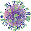
Schematic illustration of the honeycomb-shaped designer DNA nanostructure (HC-DDN)-enabled antiviral platform. The HC-DDN construct hosts antiviral agents, such as nanobodies (SD38–SD36, shown as an example), into trimeric patterns that promote multivalent interactions with hemagglutinin proteins on the influenza A virus (IAV) surface. This multivalent arrangement enables broad-spectrum viral neutralization and significantly improves cell viability in both murine and porcine models. (Image: Reproduced from DOI:10.1002/advs.202513710, CC BY) (click on image to enlarge)
The team selected a dual nanobody that targets two conserved regions of hemagglutinin. They attached a short DNA handle to each nanobody through a site-specific chemical step. That handle allows the nanobody to snap into place at the scaffold’s anchors. When the researchers positioned these nanobodies in six clusters of three across the honeycomb framework, they formed the construct known as HC Nb.
They then created HC Apt using a DNA aptamer called UHA 2. Aptamers are short strands of DNA or RNA that fold into stable shapes that recognize specific molecular surfaces. UHA 2 binds the head region of hemagglutinin and has shown activity against several influenza A subtypes. The researchers anchored UHA 2 strands in the same trimeric pattern used for the nanobodies. They also used a fluorescent aptamer that binds a different part of hemagglutinin only to measure viral entry during experiments, not as a treatment.
To determine whether spatial organization matters, the team compared free binders with scaffolded binders. They measured how strongly each version attaches to intact influenza virions. The scaffold changed the outcome sharply. Nanobodies on the DNA framework bound with roughly a thousand times greater apparent strength than nanobodies alone. Aptamers on the scaffold showed similar improvement compared to their free counterparts. These gains did not result from changes to the molecules themselves. The scaffold forces several binders to engage several hemagglutinin trimers at the same time. Each contact stabilizes the others. The virus struggles to break free.
Binding alone does not ensure real protection, so the researchers tested infection in cell culture. They exposed kidney epithelial cells, a standard model for influenza studies, to virus that had been mixed with either free nanobody or HC Nb. They kept the total amount of nanobody equal in both conditions.
After one hour, HC-Nb blocked viral entry far more effectively. In tests with a laboratory strain of H1N1, cells treated with HC-Nb showed infection levels near those of uninfected controls. Free nanobody reduced infection but still left a noticeable share of infected cells. H3N2 produced the same outcome. The scaffolded aptamer also outperformed the free aptamer, although its effect was less pronounced than the nanobody construct.
Over a full day, these differences grew. Influenza replicates quickly, and even small pockets of infection can spread and destroy healthy cells. Untreated cultures showed visible loss of viability. Virus pretreated with HC-Nb left most cells intact and functioning. Free nanobody protected some cells but failed to prevent broader damage. The scaffolded aptamer also preserved more cells than unstructured aptamer, but nanobody-based constructs remained visibly more effective.
The team extended the tests to porcine epithelial cells infected with influenza strains adapted to pigs. These systems reflect human infection more closely because pigs serve as natural hosts for influenza A virus. The scaffolded nanobody again blocked viral entry at low concentrations and preserved a large fraction of cells. Free nanobody struggled to maintain this protection even when the total number of nanobody molecules matched.
The researchers then examined whether the scaffold itself contributes to performance. They compared HC-Nb to gold nanoparticles coated with nanobodies. The nanoparticles improved antiviral activity but showed signs of cellular stress and reduced viability over time. The DNA scaffold did not show detectable toxicity in similar conditions. Measurements that track internal stress signals supported this observation. Scaffolded constructs reduced virus-induced stress more effectively than unstructured binders.
This work shows that antiviral effectiveness does not depend solely on the strength of one molecule. It also depends on how antiviral agents occupy space relative to viral proteins. The DNA scaffold does not change the chemistry of nanobodies or aptamers. It reorganizes them into trimeric clusters that mirror the pattern of hemagglutinin trimers. That alignment increases binding, reduces infection at lower doses, and preserves cells across multiple influenza A subtypes in two species.
The authors note that questions about immune response, delivery, and performance in living organisms remain. The scaffold is modular, so researchers could attach other antiviral agents using the same approach. The study illustrates how antiviral strategies may evolve toward structures that meet viruses on their own geometric terms rather than as isolated molecules drifting in solution.
For authors and communications departmentsclick to open
Lay summary
Prefilled posts
ORCID information
Xing Wang (University of Illinois at Urbana-Champaign)
, 0000-0001-9930-3287 corresponding author
Saurabh Umrao (University of Illinois at Urbana-Champaign)
, 0000-0002-9735-8062 first author
Nanowerk Newsletter
Get our Nanotechnology Spotlight updates to your inbox!
Thank you!
You have successfully joined our subscriber list.
Become a Spotlight guest author! Join our large and growing group of guest contributors. Have you just published a scientific paper or have other exciting developments to share with the nanotechnology community? Here is how to publish on nanowerk.com.