Mitochondria isolated from onions entered lung immune cells in mice and restored their energy balance, revealing an unexpected way plant-derived organelles can regulate inflammation inside mammalian tissue.
(Nanowerk Spotlight) Acute lung injury is a rapid and severe loss of lung function that follows major infection, trauma, or exposure to bacterial toxins. The condition develops when immune cells release large amounts of reactive molecules that injure the very tissue they are meant to protect.
There is still no targeted molecular therapy; treatment focuses on oxygen support and ventilation while the inflammation resolves on its own. Understanding why immune activity turns destructive and how to control that switch without impairing defense has been a central challenge in pulmonary biology. Evidence increasingly points to mitochondria, the small organelles that generate energy within cells, as critical to that process.
Mitochondria do more than produce energy. They shape how immune cells sense danger, how they generate oxidative molecules, and how they decide when to shift from attack to repair. When mitochondrial function is disrupted, inflammation intensifies and tissue damage spreads.
Laboratory studies have shown that transferring healthy mitochondria from donor cells can restore metabolism and reduce injury in animal models. Yet such approaches remain limited by practical and immunological barriers. Mammalian mitochondria are difficult to isolate in large quantities, degrade quickly, and can trigger immune rejection.
Researchers are therefore testing a different source of mitochondria: edible plants. Plant mitochondria share many structural and biochemical features with those in animals and are inherently biocompatible because they are part of the human diet. A study published in Advanced Science (“Onion‐Mitochondria Inhibit Lipopolysaccharide‐Induced Acute Lung Injury by Shaping Lung Macrophage Mitochondrial Function”) investigates whether mitochondria extracted from onion can travel from the digestive tract to the lungs and influence immune cell metabolism during acute inflammatory stress.
The study by researchers at the University of Louisville in Kentucky explores this idea in detail. The research focuses on a mouse model of lung inflammation caused by lipopolysaccharide, a component of bacterial cell walls that provokes a strong immune response. This model reliably reproduces many features of human inflammatory lung injury, including tissue swelling and overproduction of cytokines, the signaling molecules that coordinate immune activity.
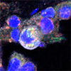
Fusion of onion-derived mitochondria (O-Mit) and macrophage mitochondria (M-Mit). A) Schematic illustration of the fusion between O-Mit and macrophage mitochondria (M-Mit) after being taken up by macrophages. Acute lung injury (ALI) mice were gavaged with DiO-labeled O-Mit. Lung tissue from ALI mice was frozen-sectioned and stained with anti-F4/80 to label macrophages, anti-TOM-20 to label M-Mit, and DAPI to label nuclei. Confocal microscopy revealed that O-Mit was taken up by macrophages in the lung tissue of ALI mice and fused with M-Mit. (400× magnification, scale bars, 200 μm). (click on image to enlarge)
The scientists compared mitochondria purified from onion, soybean, and garlic to assess their biochemical properties. Onion mitochondria contained more of a lipid called phosphatidic acid, which helps form membranes, and showed higher activity in an enzyme system known as complex I. Complex I initiates the mitochondrial energy chain that produces adenosine triphosphate, the main cellular energy currency. These traits distinguished onion mitochondria from the other plant sources.
When mice received oral doses of onion mitochondria after exposure to lipopolysaccharide, several measures of inflammation improved. Body temperature returned toward normal, the amount of fluid retained in the lungs decreased, and pro-inflammatory cytokines such as interleukin 1 beta, interferon gamma, and interleukin 6 declined. The anti-inflammatory cytokine interleukin 10 increased. Microscopic examination showed less cell infiltration and thinner alveolar walls, signs that the tissue was recovering structural stability.
Tracking experiments using fluorescent labels showed that onion mitochondria entered the bloodstream within thirty minutes of ingestion and reached peak levels two hours later. They accumulated most strongly in the lungs, particularly in animals that had been given lipopolysaccharide. The size of the mitochondria influenced where they settled. Larger ones, about two micrometers across, were more likely to remain in the lungs, while smaller ones tended to move into the liver. The inflamed state of the lung also appeared to enhance mitochondrial accumulation, probably because inflammation increases the permeability of blood vessels.
In lung tissue, most of the labeled mitochondria were found inside macrophages. These immune cells are central to inflammatory responses, releasing cytokines and clearing debris from injured tissue. When macrophages were experimentally depleted, the mitochondrial signal in the lung dropped sharply. In cultured macrophages, uptake increased when the cells were exposed to lipopolysaccharide, indicating that the inflammatory state makes them more receptive to engulfing foreign material.
Further work identified how this uptake occurs. Proteomic analysis of the macrophage membrane revealed a receptor protein called complement C3b C4b receptor 1 like. Blocking this receptor prevented the cells from internalizing onion mitochondria. The receptor recognizes phosphatidic acid, the lipid that is abundant in onion mitochondria.
Artificial vesicles made from onion phosphatidic acid were taken up efficiently by macrophages, whereas those made from other lipids were not. Removing phosphatidic acid from the mitochondrial membranes reduced uptake from most cells to very few. This pairing between the lipid and the receptor provides a molecular explanation for how onion mitochondria are selectively drawn into macrophages.
Inside the macrophages, the plant organelles fused with the cells’ own mitochondria. Imaging showed overlapping signals between the two, and chemical analysis found plant specific lipids inside the host mitochondrial fraction. When fusion was blocked, or when the onion mitochondria were damaged to disrupt their electrical potential, both fusion and the anti-inflammatory effect disappeared. This indicates that intact, functional plant mitochondria are necessary for both processes.
The fusion restored normal mitochondrial behavior. In inflamed macrophages, mitochondria usually fragment and generate excess reactive oxygen, leading to further stress. After treatment with onion mitochondria, this pattern reversed. Reactive oxygen levels dropped, antioxidant balance improved, and the mitochondrial network became long and interconnected instead of broken and punctate. Measurements of oxygen use showed that the cells shifted back to efficient energy production rather than the less efficient glycolytic mode that dominates during inflammation.
To determine what in the onion mitochondria caused these changes, the researchers analyzed their small-molecule content and identified one compound of particular interest: methyl 3,4 dihydroxybenzoate. When this compound was administered on its own, it reproduced the beneficial effects of the mitochondria. When it was removed from the mitochondrial preparation, those effects disappeared.
Methyl 3,4 dihydroxybenzoate acts on a mitochondrial gene called ND1, which encodes one subunit of complex I. The compound binds directly to ND1 DNA and lowers its expression through a chemical modification known as DNA methylation. Reducing ND1 activity decreased the production of reactive oxygen, restored the balance between oxidized and reduced cellular molecules, and reduced phosphorylation of DRP1, a protein that promotes mitochondrial splitting. The mitochondria in treated cells appeared elongated and interconnected, consistent with restored fusion.
Together, these results outline a complete mechanism. Onion mitochondria survive digestion, enter the bloodstream, and accumulate in the lungs. They attach to macrophages through a specific lipid receptor, fuse with the cells’ mitochondria, and deliver a small molecule that fine tunes mitochondrial gene activity. The outcome is less oxidative stress, fewer inflammatory cytokines, and improved lung structure and function in a standard model of acute injury.
The findings make biological sense. The lung’s narrow capillaries can physically trap particles of this size, and inflammation makes blood vessels more permeable, helping mitochondria reach the tissue. Because plant material is routinely consumed, the risk of immune rejection is low. The defined interaction between phosphatidic acid and its receptor provides a potential handle for designing mitochondria from different sources to target other tissues. The study also reported no detectable toxicity in the liver or reduction in cell growth at the tested doses.
Several questions remain. The lipopolysaccharide model represents only one type of lung injury, and the route by which plant mitochondria cross the intestinal barrier remains to be defined. Different plant species contain distinct lipid and metabolite profiles that could alter where their mitochondria go and what they do. Understanding those differences will be important for any therapeutic use. The study also raises broader questions about how mitochondrial gene regulation might be influenced by dietary compounds.
Within its defined scope, the research provides a coherent proof of concept. It shows that edible plant mitochondria can reach mammalian tissues, integrate with immune cells, and adjust how those cells manage energy and stress. By connecting plant organelles to mammalian inflammation, the study introduces a new way to think about how food components can interact with cellular machinery at the most fundamental level.
For authors and communications departmentsclick to open
Lay summary
Prefilled posts
ORCID information
Yun Teng (University of Louisville School of Medicine)
, 0000-0001-6428-2551 corresponding author
Huang-Ge Zhang (University of Louisville School of Medicine)
, 0000-0001-9665-9202 corresponding author
Nanowerk Newsletter
Get our Nanotechnology Spotlight updates to your inbox!
Thank you!
You have successfully joined our subscriber list.
Become a Spotlight guest author! Join our large and growing group of guest contributors. Have you just published a scientific paper or have other exciting developments to share with the nanotechnology community? Here is how to publish on nanowerk.com.