A magnetic microrobot navigates lung airways, drills microlesions, gathers biomarkers, and rapidly classifies cancer tissue with Raman sensing and machine learning in a single integrated workflow.
(Nanowerk Spotlight) A biopsy remains one of medicine’s most definitive tools. By directly examining tissue, doctors can confirm the presence of disease and determine how best to treat it.
In the lungs, this procedure has persistent limitations. Lesions often form in delicate, narrow airways where conventional instruments cannot reach. Standard bronchoscopes and forceps are too large to extract useful samples from tiny nodules, while needle biopsies guided by imaging carry risks of bleeding or collapsed lung.
Even when tissue can be collected, the sample is sometimes too small or degraded to allow precise analysis. To overcome this, researchers have developed thinner bronchoscopes that can extend deeper, catheters able to curve into side branches, and robotic systems that stabilize needle placement under imaging. These advances extend access but still fail in the smallest airways. Flexible scopes lack the stability to extract intact tissue, and image-guided needles can miss lesions that move with each breath. Many patients face repeat procedures or remain without a firm diagnosis.
Recent work in miniature robotics and molecular sensing suggests another path. Micromachines smaller than a grain of rice can be fabricated with magnetic coatings and geometries that let them screw through liquid-filled passages.
Optical methods such as surface-enhanced Raman spectroscopy, which uses nanostructured metal to magnify the light scattered by molecules, now detect distinct chemical fingerprints from only a trace of material. Combined with machine learning, these signals can be classified into diagnostic categories within minutes.
Schematic of the multifunctional microrobotic platform for targeted biopsy and SERS-based diagnosis of deep-seated microlesions. A conical helix structuredmicrorobot consisting of imaging barium sulfate agents,magnetic nickel, and biocompatible platinum surface layers, as well as abundant gold nanospikes at the tip, is developed as the biopsy and biosensing tool. The procedures mainly include five stages, i.e., the tracheoscope intervention and saline injection followed by deployment of microrobot (I), magnetic navigation to the targeted tracheal lesions (II), drilling-enabled tissue and biomarker sampling at the target (III), moving back and retrieval of microrobot (IV), and finally cancer diagnosis via SERS sensing. The real-time control of microrobot in lung was realized under the actuation of a rotating magnetic field and the tracking of tracheoscopy and fluoroscopy. Hydrogen peroxide (the biomarker) concentration can be quantified by modifying the Au nanospikes at microrobot tip with Raman reporter molecule, while various types of sampled lung tissues can be recognized by deep learning-reinforced Raman detection. (Image: Reprinted with permission from Wiley-VCH Verlag) (click on image to enlarge)
The microrobot is a conical helix about the size of a pinhead. Its narrowing shape reduces the chance of scraping airway walls during movement. It is built with micro-scale 3D printing and coated with nickel for magnetic response and platinum for biocompatibility. At the very tip, gold nanospikes are grown in a controlled chemical process.
These nanospikes act both as a scraper to collect material and as an enhancer of Raman signals when illuminated by a laser. Tests confirmed signal amplification on the order of one hundred sixty thousand, sufficient for detecting molecules at very low concentrations.
Motion is driven by a rotating magnetic field. When the external magnet spins, the helical body advances like a screw through fluid.
In test channels, the robot reached more than one millimeter per second and resisted both forward and reverse flow. Fluid simulations confirmed that the spiral geometry reduces drag and stabilizes motion. By adjusting the orientation of the magnetic field, operators can either press the tip firmly against a surface for drilling or guide it forward in a controlled glide. This flexibility is essential for reaching lesions located at different sides of the airway.
The device was tested in progressively more complex settings. It was first navigated through a 3D-printed airway model filled with saline, then through ex vivo pig lungs, and finally in a live rabbit.
In these tests, the robot was deployed through a catheter introduced by tracheoscope. After release into the fluid-filled airway, it was controlled by an external magnet mounted on a robotic arm. Its position was tracked using fluoroscopy and endoscopy.
In both the pig lungs and the rabbit, the microrobot reached distal branches, made contact with a target, collected samples, and was retrieved successfully. No adverse effects were reported. To ensure visibility under X-ray and strong magnetic response, barium sulfate and neodymium-iron-boron particles were embedded into the body during fabrication.
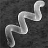
SEM images of microrobot, and the magnified view of its tip that shows the growth and distribution of Au nanospikes. (Image: Reprinted with permission from Wiley-VCH Verlag)
The device also functions as a chemical sensor. Raman spectroscopy measures scattered light that reveals molecular vibrations. Because these signals are normally weak, the nanospikes at the tip serve as amplifiers through a process called surface-enhanced Raman scattering, or SERS.
To test this capability, the researchers coated the spikes with a probe molecule that reacts with hydrogen peroxide, a compound often elevated in tumor environments. When exposed to peroxide, the probe’s Raman spectrum changes, producing a new peak.
In a lung model, the robot was directed to an airway injected with peroxide. After retrieval, the spectrum showed the expected peak, and the measured concentration closely matched the known value of the injected solution. A control robot deployed to a peroxide-free region showed no peak, demonstrating specificity of detection.
The microrobot can also collect cellular material. In cultured cell layers, the tip was driven across colonies, leaving cleared patches.
Microscopy showed fragments of cells lodged among the nanospikes, and protein assays confirmed nanogram-scale amounts were collected. Raman spectra from these samples revealed cell-associated features not present in control robots. Simulations estimated that the sampling force was sufficient to dislodge cells but not strong enough to detach the nanospikes, ensuring structural stability.
To test diagnostic use, the researchers examined human lung tissue sections representing normal tissue and four cancer types: small cell carcinoma, large cell carcinoma, squamous cell carcinoma, and adenocarcinoma.
The robot tip drilled lightly into the tissues to load trace samples. Spectra were then acquired from these samples. Although average spectra across tissue types looked similar, a computational model identified consistent distinctions.
The model used was a convolutional neural network, a type of pattern recognition system that learns by scanning data for recurring features. It was trained on most of the collected spectra and tested on the remainder. Overall classification accuracy reached about 94 percent, with near-perfect results for the cancer tissues and somewhat lower accuracy for normal tissue. Once trained, the model delivered classifications within about three minutes.
The design integrates several elements to combine navigation and sensing. Barium sulfate provides visibility under X-ray. Magnetic coatings and embedded particles allow precise steering from outside the body. The conical helix centers the device in the airway, protecting surrounding tissue. The gold nanospikes act both as the contact surface for sampling and as the amplification surface for Raman detection, so the same site is used for both collection and analysis. Each choice addresses a barrier that has hindered earlier attempts at minimally invasive diagnosis.
The work remains at the stage of feasibility testing. The navigation trials were conducted in pig lungs and rabbits, and peroxide was used as a proxy for more complex tumor markers.
The researchers identify clear next steps. Protective coatings could prevent contamination before sampling. Different probe molecules could be attached to detect a broader range of biomarkers. Drug payloads might be incorporated for combined sampling and therapy. Larger animal studies and tighter integration with medical imaging will be needed before any clinical trials in humans.
The study demonstrates a device that can reach beyond the limits of bronchoscopes and needles, collect samples from otherwise inaccessible sites, and analyze those samples immediately.
By merging microrobotics, magnetic actuation, nanostructured sensing, and computational classification, the work in Advanced Materials shows a way to shorten the path from detecting a suspicious lung lesion to identifying its nature with speed and precision.
For authors and communications departmentsclick to open
Lay summary
Prefilled posts
Nanowerk Newsletter
Get our Nanotechnology Spotlight updates to your inbox!
Thank you!
You have successfully joined our subscriber list.
Become a Spotlight guest author! Join our large and growing group of guest contributors. Have you just published a scientific paper or have other exciting developments to share with the nanotechnology community? Here is how to publish on nanowerk.com.