A multimodal imaging study combines cryo-X-ray nanotomography and super-resolution fluorescence microscopy to reveal how protein-coated nanoparticles behave in cells.
(Nanowerk Spotlight) The biological effects of engineered nanoparticles do not arise solely from their design but also from how they interact with their environment once inside the body. The moment nanoparticles come into contact with blood, mucus, or intracellular fluid, they are rapidly covered by a layer of proteins and other biomolecules. This layer, known as the protein corona, alters nearly every aspect of nanoparticle behavior—from their physical stability and circulation time to how they are recognized, absorbed, and processed by cells.
Despite extensive interest in nanoparticle-based therapies and diagnostics, a key obstacle remains: scientists cannot fully predict how nanoparticles will behave inside cells over time, especially once the corona has formed.
This problem has slowed progress in clinical translation. Most laboratory studies rely on fixed samples, where nanoparticles are introduced to cells, and after a set period, the cells are chemically preserved and imaged. While useful, this approach cannot distinguish early from late stages of nanoparticle uptake. Nor can it reveal the dynamic transitions nanoparticles undergo once inside the cell. Without detailed knowledge of how particles move, cluster, or degrade over time, researchers lack the information needed to optimize design features for safety, specificity, or therapeutic action.
Advances in imaging now make it possible to follow nanoparticles inside intact cells across multiple time points. Cryogenic soft X-ray tomography, for example, preserves hydrated samples in their natural state and generates high-resolution three-dimensional reconstructions without requiring contrast agents or disruptive sample preparation. When combined with super-resolution fluorescence microscopy and correlative workflows, it becomes possible to match structural and molecular information with high temporal and spatial resolution. These capabilities are essential for building a more complete picture of how cells internalize nanoparticles and what happens to them afterwards.
Their method addressed key limitations in earlier studies, particularly the inability to track internalization events with clear time resolution. By labeling the nanoparticles with fluorescent markers and combining cryo-fixation at defined time points with a suite of imaging methods, the team created a framework that preserved both structural and chronological detail.
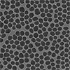
Nanoparticle and protein corona characterization. a) SiNP TEM micrograph and b) its corresponding size distribution (N > 200). c) Illustration depicting the chemical surfaces of SiNP, SiNP-S, and SiNP-P. d) Mass of adsorbed proteins per mass of nanoparticles after incubation with BSA and FBS (N = 3). e) DH of SiNP in cell culture media without protein, and with the addition of 10 g L−1 BSA or 10% FBS. f) DH by NNLS method of SiNP, SiNP-S, and SiNP-P in cell culture media without protein, and with the addition of BSA or FBS. g) Cryo-TEM image of SiNP in cell culture media supplemented with 10% FBS. h) Illustration demonstrating the colloidal stability of the nanoparticles in the presence of BSA or FBS. (Image: reprinted with permission by Wiley-VCH Verlag) (click on image to enlarge)
The team began by synthesizing uniform silica nanoparticles with an average diameter of 55 nanometers. These particles were either left unmodified (SiNP) or functionalized with polyethylene glycol (PEG) or sulfobetaine silane (SBS), both of which are known to reduce protein adsorption. The researchers then incubated the particles in media containing either purified bovine serum albumin (BSA) or full fetal bovine serum (FBS), allowing protein coronas to form. As expected, the unmodified SiNPs adsorbed significantly more protein—around ten times more—than their functionalized counterparts. The PEG- and SBS-coated particles still formed coronas, but of much lower density.
Tests in NIH-3T3 fibroblast cells showed that protein coronas influenced both nanoparticle uptake and cell viability. Uncoated SiNPs caused significant toxicity in serum-free media, while protein-coated and functionalized particles did not. Uptake efficiency also varied by corona type: BSA-coated SiNPs entered cells more readily than FBS-coated ones, even though BSA represents only a fraction of the total protein in FBS. This suggests that components of the more complex FBS corona may interfere with pathways responsible for cellular uptake.
To monitor what happened after the particles entered the cells, the team exposed fibroblasts to nanoparticles for one hour, washed away any particles that remained outside the cell, and then cryo-fixed the cells at 0, 2, or 24 hours later. Widefield fluorescence microscopy showed a shift in the location of nanoparticles over time—from the outer cytoplasmic region toward the area surrounding the nucleus. Electron microscopy revealed that all internalized nanoparticles were enclosed in vesicles, with no evidence of free particles in the cytosol. This indicates that endocytosis was the primary uptake mechanism and that the protein corona may prevent cytosolic release, reducing the likelihood of damage to cellular components.
The use of cryogenic soft X-ray tomography provided three-dimensional views of intact cells, showing that nanoparticles resided within membrane-bound vesicles whose location and size changed over time. Initially, these vesicles were smaller and closer to the cell membrane. After 24 hours, they were larger and concentrated around the nucleus. Quantitative measurements showed that vesicle diameter increased from about 0.7–0.8 micrometers to 1.2 micrometers. This expansion is consistent with vesicle fusion events, a hallmark of endosomal maturation.
To further investigate the identity of the vesicles, the researchers used cryogenic structured illumination microscopy to label acidic compartments, such as late endosomes and lysosomes. Correlating these results with the X-ray data revealed that some vesicles containing nanoparticles had acidic interiors, while others did not. This finding indicates that nanoparticles passed through different stages of the endosomal system. The presence of particles in both early and late vesicles 24 hours after uptake also suggests that clearance or degradation of these particles was minimal over that time frame.
Electron microscopy provided additional evidence for vesicle maturation. Distinct vesicle types were identified based on shape and internal structure, including early endosomes, late endosomes with internal compartments, and larger vesicles likely to be endolysosomes. The researchers also observed apparent fusion events between vesicles, reinforcing the interpretation that particle-containing compartments undergo dynamic reorganization during intracellular trafficking.
To quantify localization changes, the team calculated the distance from each vesicle to the nucleus and the membrane. They found that, immediately after exposure, most vesicles were near the cell edge. After 24 hours, most were closer to the nucleus. This trend was more pronounced for BSA-coated particles than for those coated in FBS, supporting earlier observations that corona composition affects not just uptake but also intracellular routing.
Overall, the study shows that nanoparticles do not remain static after entering a cell. Instead, they are actively moved through a series of vesicles that grow, merge, and change in acidity over time. The protein corona plays a decisive role in shaping how quickly and through which paths this process unfolds. The multimodal imaging approach developed here allows researchers to observe these changes with precision that was previously difficult to achieve.
These insights have important implications for nanoparticle design. Understanding how different coronas influence uptake, retention, and vesicle trafficking can inform strategies to control drug release timing, avoid lysosomal degradation, or target specific subcellular regions. The ability to track particles in intact, hydrated cells over defined time intervals provides a strong platform for future studies. By applying these methods to a wider range of particle types and biological contexts, researchers can develop clearer rules for designing nanomaterials that behave predictably inside living systems.
Get our Nanotechnology Spotlight updates to your inbox!
Thank you!
You have successfully joined our subscriber list.
Become a Spotlight guest author! Join our large and growing group of guest contributors. Have you just published a scientific paper or have other exciting developments to share with the nanotechnology community? Here is how to publish on nanowerk.com.