Researchers create self-assembling protein nanofibers with intrinsic photoconductivity, enabling biointegrated devices responsive to near-infrared light.
(Nanowerk Spotlight) Protein-based nanomaterials that respond to light are emerging as candidates for integrating biological compatibility with programmable electronic behavior. Their appeal lies in the possibility of designing flexible, degradable components that can function in aqueous and physiological environments—something difficult to achieve with traditional semiconductors.
Existing photoconductive materials, including inorganic semiconductors and conjugated polymers, typically require rigid crystalline structures or synthetic modification to function. Their lack of tunability, limited compatibility with biological systems, and dependence on harsh fabrication methods restrict their use in bioelectronics and light-controlled therapeutics.
Researchers have long explored conductive proteins inspired by natural examples such as the pili of Geobacter sulfurreducens, which show conductivity due to stacking of aromatic amino acids—chemical groups containing planar carbon ring structures. These findings led to the development of engineered microbial systems and peptide-based nanostructures capable of transporting charge. Some short aromatic peptides synthesized with fluorenylmethyloxycarbonyl chemistry have shown low-level conduction, in the range of 10⁻¹⁰ to 10⁻⁹ siemens per centimeter.
But while these structures demonstrate electron transport, they do not respond to light. Other efforts to introduce photosensitivity into biomolecules have relied on synthetic attachments such as photoswitchable dyes or conjugated oligomers. These modifications, while functional, introduce complexity and often compromise biocompatibility.
A critical missing step has been the design of fully genetically encoded, self-assembling protein systems that are both conductive and photoresponsive. Such systems would eliminate the need for chemical conjugation and allow bottom-up construction of optoelectronic materials from sequence alone.
In a new study published in Advanced Functional Materials (“Protein‐Engineered Photoresponsive Conductive Nanofibers”), researchers at New York University present a protein fiber architecture that meets this challenge. Using rational sequence design and computational modeling, they created two coiled-coil protein nanofibers—named CHAF and L-CHAF—that exhibit light-induced electrical response in the near-infrared region.
The team constructed CHAF by placing phenylalanine residues—aromatic amino acids—at structurally strategic positions within a coiled-coil scaffold. These positions correspond to the “a” and “d” slots of the heptad repeat pattern typical of coiled-coils, where hydrophobic residues pack into the fiber’s core. A control version, L-CHAF, was designed with leucine—non-aromatic but hydrophobic—in place of phenylalanine. Both sequences were expressed in E. coli, purified, and characterized structurally and functionally.
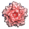
CHAF/L-CHAF protein sequence labeled by helical wheel positions a–g. CHAF protein possesses F in the a and d positions whereas L-CHAF protein possess L in the a and d positions. Cartoon of CHAF protein is displayed as a ribbon diagram with phenylalanines highlighted as red sticks and as an electrostatic potential map from N- to C-terminus. Positive blue and negative red patches represent a scale of −10 to 10 kbT. Schematic represents the proposed self-assembly by predominant end-to-end stacking of coiled-coils. (Image: reprinted with permission by Wiley-VCH Verlag)
Electron microscopy confirmed that the two proteins self-assemble into nanoscale fibers with diameters around 24 nanometers. Both remained stable in aqueous buffer and exhibited expected structural features. Spectroscopic measurements showed that CHAF was less helical and more disordered than L-CHAF, likely due to the bulkier aromatic rings disrupting the coiled-coil packing. This disruption reduced structural rigidity but created an internal environment more conducive to stacking interactions between aromatic side chains.
Despite these differences, both CHAF and L-CHAF displayed measurable conductivity and photoresponse. The researchers fabricated simple photodetector devices by drop-casting the fibers onto gold electrodes. When illuminated with light at 808 nanometers, both materials generated photocurrents, with L-CHAF showing higher baseline current and stronger response. The device response scaled with light intensity and remained stable across multiple on/off cycles during testing. Importantly, the devices worked without requiring additional conductive additives or synthetic modifications. Their active elements were entirely protein-based.
The mechanism behind this photoresponsivity appears to involve excimer formation. Excimers are short-lived complexes formed when an aromatic molecule in an excited electronic state interacts with a nearby ground-state partner. In the case of CHAF, phenylalanine residues form the excimer; for L-CHAF, histidine residues, which also possess aromatic rings, likely play a similar role.
The authors suggest that when voltage is applied across the protein film, redox-active side chains at the electrodes undergo electron transfer, creating the excited-state precursors for excimer formation. Once formed, these excimers absorb light in the visible and near-infrared range, enabling the observed photoconductivity.
Molecular dynamics simulations and quantum mechanical calculations support this interpretation. Time-dependent density functional theory (TDDFT) showed that phenylalanine side chains within the fiber core can absorb energy and form delocalized excited states, with excitation energies matching the wavelengths used in the experiment.
The same modeling revealed that charge transport likely occurs via electron hopping—where electrons jump between localized sites along and between protein chains. This process is assisted by aromatic stacking and the structural order of the fibers, which helps align these sites for more efficient transport.
The higher photocurrent in L-CHAF is explained by its increased structural order. The more tightly packed helices promote inter-chain connectivity and reduce charge trapping, which often slows down or dampens photoresponse. Though lacking the aromatic core of CHAF, the histidine residues in L-CHAF still support pi-stacking interactions and redox activity, offering an alternative conduction pathway that benefits from the more ordered molecular framework.
What sets this work apart is its complete reliance on encoded protein sequence to direct material behavior. These fibers are not chemically modified after synthesis. Their photoconductivity arises from properties intrinsic to the amino acid side chains and their arrangement in the coiled-coil scaffold. This design approach allows for modularity, tunability, and potential integration into biologically relevant environments.
Possible applications include bioresorbable sensors that respond to light, implantable devices that communicate optically, and programmable scaffolds for drug release triggered by infrared illumination. Unlike traditional photodetectors, these protein-based systems could be used in soft tissue or aqueous conditions without toxicity. Their fabrication requires no high-temperature processing or complex lithography—just gene expression, purification, and self-assembly.
The study illustrates that single-domain proteins, when carefully designed, can exhibit electronic properties typically associated with synthetic semiconductors. It also opens a path to design photodetectors, light-responsive actuators, or hybrid systems using engineered proteins alone. Future iterations may include engineered redox centers, higher-order assemblies, or integration with other biological components to expand function.
By combining precise sequence control, supramolecular design, and photophysical modeling, this work demonstrates a platform where photoresponsive behavior is an emergent property of protein architecture. It lays the foundation for constructing optoelectronic devices that are biodegradable, biointegrated, and genetically programmable.
Get our Nanotechnology Spotlight updates to your inbox!
Thank you!
You have successfully joined our subscriber list.
Become a Spotlight guest author! Join our large and growing group of guest contributors. Have you just published a scientific paper or have other exciting developments to share with the nanotechnology community? Here is how to publish on nanowerk.com.