Microscale 3D printed arches guide cells to form natural self-bonds, improving how soft tissue attaches to medical implants without added proteins.
(Nanowerk Spotlight) Efforts to engineer reliable connections between soft living tissue and synthetic materials have struggled with persistent instability at the interface. When a medical implant crosses the skin or interfaces with soft internal tissues, a secure and long-lasting bond is often difficult to achieve.
These interfaces—found in devices like dental screws, bone-anchored prosthetics, catheters, and neural implants—are vulnerable to a range of complications, including infection, inflammation, and mechanical failure. The biological response at these junctions is typically unpredictable, and even modest misalignment or degradation can provoke tissue rejection or device encapsulation.
Traditional solutions rely heavily on encouraging cells to attach through integrins, proteins that mediate adhesion to the extracellular matrix. This approach has led to the widespread use of biomaterial coatings such as fibronectin, collagen, or short peptides that mimic matrix-binding sites. While these coatings can promote temporary attachment, they are susceptible to enzymatic degradation and remodeling. Their short lifespan and inability to mimic the dynamic architecture of living tissue limit their long-term effectiveness, particularly for implants that remain in the body for extended periods.
Another critical class of adhesion proteins—cadherins—has attracted growing interest in the field. These proteins mediate cell-to-cell adhesion through calcium-dependent binding and play central roles in maintaining tissue structure, regulating cellular movement, and transmitting mechanical signals. Unlike integrins, cadherins bind cells directly to one another through a highly specific and mechanically responsive interface.
Although some researchers have tried to use cadherin-mimetic coatings or recombinant cadherin proteins to promote attachment, these strategies also suffer from surface degradation and fail to replicate the full complexity of natural cell-cell interactions.
To bypass these constraints, a research team from Princeton University developed a new strategy. Rather than mimic cadherin binding externally, they engineered a three-dimensional microstructure that encourages a cell to form a cadherin-mediated junction with itself. This phenomenon, known as cellular self-adhesion, occurs naturally in a few biological systems—most notably in certain neural and vascular tissues where a single cell loops around and binds to itself. The researchers designed a synthetic surface that prompts this behavior using a nanoscale tunnel-like structure, enabling cells to engage their cadherin machinery in a controlled and stable manner.
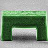
In a study published in Advanced Materials (“Engineering Cellular Self‐Adhesions Inside 3D Printed Micro‐Arches to Enhance Cell:Biomaterial Attachment”), the team introduced Self-Adhesion Tunnels (SATs)—subcellular-scale micro-arches fabricated directly onto glass surfaces using two-photon polymerization, a high-resolution 3D printing technique. These SATs are small enough to fit within a single cell’s footprint, creating a confined space that brings opposing regions of the cell membrane into close contact. This geometry encourages the formation of E-cadherin-mediated junctions inside the tunnel, essentially creating a cell–material interface built from the cell’s own internal adhesive system.
Fabrication and characterization of 3D printed Self-Adhesion Tunnel (SAT) arrays. a) Fabrication process for SATs using 2-photon polymerization. b) Cartoon hypothesis of SAT topology resulting in the formation of a cell self-adhesion. c) 3D render from confocal scan of a medium size SAT. d) Confocal images showing E-cadherin localization inside of the SAT cavity. e) Scanning electron micrograph (SEM) of a medium arch (scale bar = 5 μm). f) Fluorescence image in XY-plane of MDCK monolayer on an array of SATs (scale bar = 50 μm). g) SEM image of MDCK cells attached to SATs (scale bar = 10 μm). (Image: reprinted from DOI:10.1002/adma.202502425, CC BY)
The team tested their SAT structures using a kidney epithelial cell model known for forming strong cadherin junctions. They observed that cells reliably formed self-adhesions inside the tunnels, with E-cadherin and other components of the cell–cell junctional complex localizing inside the SATs. These structures included beta-catenin and F-actin, which link cadherins to the cytoskeleton and mediate force transmission, as well as desmoplakin, a protein found in mature, mechanically robust junctions. The presence of all these components suggested that the SATs do not interfere with junction maturation or signaling and instead recreate a normal adhesive architecture within a synthetic context.
To improve the efficiency of this adhesion mechanism, the researchers explored how changes in SAT geometry affected self-adhesion formation. They tested a range of sizes and shapes and found that smaller SATs produced higher frequencies of self-adhesion, likely because they required less membrane deformation. A trapezoidal version of the SAT, which maintained the tunnel shape while reducing surface area, achieved the highest efficiency—up to 93% of eligible SATs successfully induced a self-adhesion. This optimization process demonstrated that geometry alone, without any chemical coating or biological modification, can control and improve the adhesive behavior of cells.
Further analysis revealed that these self-adhesions were not only stable but mechanically similar to natural cell–cell junctions. Using a technique called fluorescence recovery after photobleaching (FRAP), the team compared how quickly cadherin proteins turned over in self-adhesions versus normal cell–cell contacts. The results showed no significant difference in recovery time, indicating that the dynamics and mechanical stability of the engineered junctions closely resemble those of their natural counterparts.
The researchers also tested whether these structures could stabilize tissue over time. Using engineered sheets of primary skin cells placed over SAT arrays, they observed that self-adhesions persisted for hours and reduced the speed of collective cell migration compared to cells grown on flat glass or control structures without tunnels. These effects suggest that SATs not only enable attachment but may also improve the mechanical integration between tissue and biomaterial surfaces. In long-term trials, self-adhesions remained functional across multiple days, and mitotic activity appeared unaffected, indicating that the structures do not disrupt normal cell behavior.
To assess whether the approach generalizes beyond a single tissue type, the researchers evaluated SAT performance in other epithelial and endothelial cell types. They observed successful self-adhesion formation in both human induced pluripotent stem cells and human endothelial cells, using E-cadherin and VE-cadherin markers, respectively. This suggests that SATs can support a range of cadherin subtypes and tissue origins, making them a flexible tool for tissue engineering.
Unlike chemical coatings, which degrade or diffuse over time, SATs offer a physically defined, protein-free structure that interacts directly with the cell’s own adhesive machinery. This strategy has several implications. It could improve the design of percutaneous implants, where the skin-device interface is a frequent failure point. It could also support the development of organoid platforms or engineered tissues that require stable, long-term adhesion in environments where conventional ECM coatings fall short.
Additionally, the use of SATs as a standardized platform could facilitate the study of cadherin dynamics under defined mechanical and spatial constraints, potentially yielding new insights into cell adhesion biology.
The work offers a blueprint for how engineered topographies can be used not just as passive substrates but as active guides for cellular behavior. By encouraging cells to anchor themselves through a natural, internally regulated process, this approach moves beyond biochemical mimicry and introduces a geometric principle for designing stable biointerfaces. While further testing will be needed in complex tissue models and in vivo systems, the ability to induce and control self-adhesions using entirely synthetic materials represents a meaningful step toward more reliable and resilient soft tissue integration.
Get our Nanotechnology Spotlight updates to your inbox!
Thank you!
You have successfully joined our subscriber list.
Become a Spotlight guest author! Join our large and growing group of guest contributors. Have you just published a scientific paper or have other exciting developments to share with the nanotechnology community? Here is how to publish on nanowerk.com.